Sodium »
PDB 3qq0-3r57 »
3r0x »
Sodium in PDB 3r0x: Crystal Structure of Selenomethionine Incorporated Apo D-Serine Deaminase From Salmonella Tyhimurium
Enzymatic activity of Crystal Structure of Selenomethionine Incorporated Apo D-Serine Deaminase From Salmonella Tyhimurium
All present enzymatic activity of Crystal Structure of Selenomethionine Incorporated Apo D-Serine Deaminase From Salmonella Tyhimurium:
4.3.1.18;
4.3.1.18;
Protein crystallography data
The structure of Crystal Structure of Selenomethionine Incorporated Apo D-Serine Deaminase From Salmonella Tyhimurium, PDB code: 3r0x
was solved by
S.R.Bharath,
B.Shveta,
H.S.Savithri,
M.R.N.Murthy,
with X-Ray Crystallography technique. A brief refinement statistics is given in the table below:
| Resolution Low / High (Å) | 36.17 / 1.93 |
| Space group | P 21 21 2 |
| Cell size a, b, c (Å), α, β, γ (°) | 56.460, 188.390, 46.590, 90.00, 90.00, 90.00 |
| R / Rfree (%) | 18.1 / 21 |
Sodium Binding Sites:
The binding sites of Sodium atom in the Crystal Structure of Selenomethionine Incorporated Apo D-Serine Deaminase From Salmonella Tyhimurium
(pdb code 3r0x). This binding sites where shown within
5.0 Angstroms radius around Sodium atom.
In total only one binding site of Sodium was determined in the Crystal Structure of Selenomethionine Incorporated Apo D-Serine Deaminase From Salmonella Tyhimurium, PDB code: 3r0x:
In total only one binding site of Sodium was determined in the Crystal Structure of Selenomethionine Incorporated Apo D-Serine Deaminase From Salmonella Tyhimurium, PDB code: 3r0x:
Sodium binding site 1 out of 1 in 3r0x
Go back to
Sodium binding site 1 out
of 1 in the Crystal Structure of Selenomethionine Incorporated Apo D-Serine Deaminase From Salmonella Tyhimurium
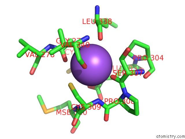
Mono view
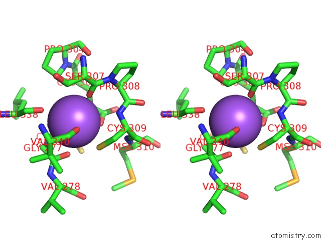
Stereo pair view
Mono view
Stereo pair view
A full contact list of Sodium with other atoms in the Na binding
site number 1 of Crystal Structure of Selenomethionine Incorporated Apo D-Serine Deaminase From Salmonella Tyhimurium within 5.0Å range:
|
Reference:
S.R.Bharath,
S.Bisht,
H.S.Savithri,
M.R.N.Murthy.
Crystal Structures of Open and Closed Forms of D-Serine Deaminase From Salmonella Typhimurium - Implications on Substrate Specificity and Catalysis Febs J. 2011.
ISSN: ISSN 1742-464X
PubMed: 21668644
DOI: 10.1111/J.1742-4658.2011.08210.X
Page generated: Mon Oct 7 12:44:02 2024
ISSN: ISSN 1742-464X
PubMed: 21668644
DOI: 10.1111/J.1742-4658.2011.08210.X
Last articles
Fe in 2YXOFe in 2YRS
Fe in 2YXC
Fe in 2YNM
Fe in 2YVJ
Fe in 2YP1
Fe in 2YU2
Fe in 2YU1
Fe in 2YQB
Fe in 2YOO