Sodium »
PDB 3q11-3qpz »
3qfh »
Sodium in PDB 3qfh: 2.05 Angstrom Resolution Crystal Structure of Epidermin Leader Peptide Processing Serine Protease (Epip) From Staphylococcus Aureus.
Protein crystallography data
The structure of 2.05 Angstrom Resolution Crystal Structure of Epidermin Leader Peptide Processing Serine Protease (Epip) From Staphylococcus Aureus., PDB code: 3qfh
was solved by
G.Minasov,
A.Halavaty,
L.Shuvalova,
I.Dubrovska,
J.Winsor,
F.Bagnoli,
F.Falugi,
M.Bottomley,
G.Grandi,
W.F.Anderson,
Center For Structuralgenomics Of Infectious Diseases (Csgid),
with X-Ray Crystallography technique. A brief refinement statistics is given in the table below:
| Resolution Low / High (Å) | 29.69 / 2.05 |
| Space group | P 1 |
| Cell size a, b, c (Å), α, β, γ (°) | 85.505, 94.696, 122.999, 89.98, 90.37, 116.79 |
| R / Rfree (%) | 17 / 21.6 |
Sodium Binding Sites:
The binding sites of Sodium atom in the 2.05 Angstrom Resolution Crystal Structure of Epidermin Leader Peptide Processing Serine Protease (Epip) From Staphylococcus Aureus.
(pdb code 3qfh). This binding sites where shown within
5.0 Angstroms radius around Sodium atom.
In total 3 binding sites of Sodium where determined in the 2.05 Angstrom Resolution Crystal Structure of Epidermin Leader Peptide Processing Serine Protease (Epip) From Staphylococcus Aureus., PDB code: 3qfh:
Jump to Sodium binding site number: 1; 2; 3;
In total 3 binding sites of Sodium where determined in the 2.05 Angstrom Resolution Crystal Structure of Epidermin Leader Peptide Processing Serine Protease (Epip) From Staphylococcus Aureus., PDB code: 3qfh:
Jump to Sodium binding site number: 1; 2; 3;
Sodium binding site 1 out of 3 in 3qfh
Go back to
Sodium binding site 1 out
of 3 in the 2.05 Angstrom Resolution Crystal Structure of Epidermin Leader Peptide Processing Serine Protease (Epip) From Staphylococcus Aureus.
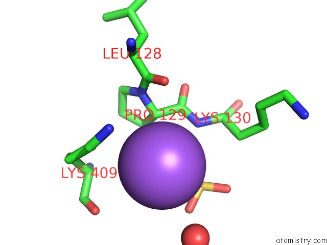
Mono view
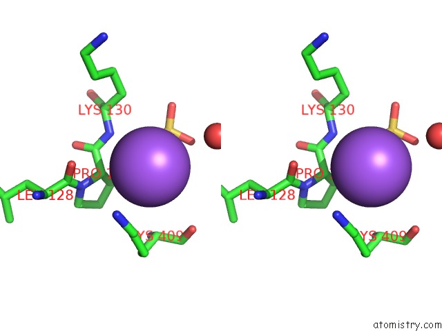
Stereo pair view
Mono view
Stereo pair view
A full contact list of Sodium with other atoms in the Na binding
site number 1 of 2.05 Angstrom Resolution Crystal Structure of Epidermin Leader Peptide Processing Serine Protease (Epip) From Staphylococcus Aureus. within 5.0Å range:
|
Sodium binding site 2 out of 3 in 3qfh
Go back to
Sodium binding site 2 out
of 3 in the 2.05 Angstrom Resolution Crystal Structure of Epidermin Leader Peptide Processing Serine Protease (Epip) From Staphylococcus Aureus.

Mono view

Stereo pair view
Mono view
Stereo pair view
A full contact list of Sodium with other atoms in the Na binding
site number 2 of 2.05 Angstrom Resolution Crystal Structure of Epidermin Leader Peptide Processing Serine Protease (Epip) From Staphylococcus Aureus. within 5.0Å range:
|
Sodium binding site 3 out of 3 in 3qfh
Go back to
Sodium binding site 3 out
of 3 in the 2.05 Angstrom Resolution Crystal Structure of Epidermin Leader Peptide Processing Serine Protease (Epip) From Staphylococcus Aureus.

Mono view

Stereo pair view
Mono view
Stereo pair view
A full contact list of Sodium with other atoms in the Na binding
site number 3 of 2.05 Angstrom Resolution Crystal Structure of Epidermin Leader Peptide Processing Serine Protease (Epip) From Staphylococcus Aureus. within 5.0Å range:
|
Reference:
G.Minasov,
A.Halavaty,
L.Shuvalova,
I.Dubrovska,
J.Winsor,
F.Bagnoli,
F.Falugi,
M.Bottomley,
G.Grandi,
W.F.Anderson.
2.05 Angstrom Resolution Crystal Structure of Epidermin Leader Peptide Processing Serine Protease (Epip) From Staphylococcus Aureus. To Be Published.
Page generated: Mon Oct 7 12:30:59 2024
Last articles
Mn in 5Z5LMn in 5Z2R
Mn in 5Z2K
Mn in 5Y87
Mn in 5YN3
Mn in 5YVS
Mn in 5YVR
Mn in 5YVM
Mn in 5YTZ
Mn in 5YTY