Sodium »
PDB 3pkk-3pzs »
3pws »
Sodium in PDB 3pws: Crystal Structure of Aspartate Beta-Semialdehide Dehydrogenase From Streptococcus Pneumoniae with 2',5'-Adenosine Diphosphate and D-2- Aminoadipate
Enzymatic activity of Crystal Structure of Aspartate Beta-Semialdehide Dehydrogenase From Streptococcus Pneumoniae with 2',5'-Adenosine Diphosphate and D-2- Aminoadipate
All present enzymatic activity of Crystal Structure of Aspartate Beta-Semialdehide Dehydrogenase From Streptococcus Pneumoniae with 2',5'-Adenosine Diphosphate and D-2- Aminoadipate:
1.2.1.11;
1.2.1.11;
Protein crystallography data
The structure of Crystal Structure of Aspartate Beta-Semialdehide Dehydrogenase From Streptococcus Pneumoniae with 2',5'-Adenosine Diphosphate and D-2- Aminoadipate, PDB code: 3pws
was solved by
A.G.Pavlovsky,
R.E.Viola,
with X-Ray Crystallography technique. A brief refinement statistics is given in the table below:
| Resolution Low / High (Å) | 36.73 / 2.00 |
| Space group | P 1 21 1 |
| Cell size a, b, c (Å), α, β, γ (°) | 60.082, 99.404, 64.612, 90.00, 101.14, 90.00 |
| R / Rfree (%) | 20.4 / 23.1 |
Sodium Binding Sites:
The binding sites of Sodium atom in the Crystal Structure of Aspartate Beta-Semialdehide Dehydrogenase From Streptococcus Pneumoniae with 2',5'-Adenosine Diphosphate and D-2- Aminoadipate
(pdb code 3pws). This binding sites where shown within
5.0 Angstroms radius around Sodium atom.
In total only one binding site of Sodium was determined in the Crystal Structure of Aspartate Beta-Semialdehide Dehydrogenase From Streptococcus Pneumoniae with 2',5'-Adenosine Diphosphate and D-2- Aminoadipate, PDB code: 3pws:
In total only one binding site of Sodium was determined in the Crystal Structure of Aspartate Beta-Semialdehide Dehydrogenase From Streptococcus Pneumoniae with 2',5'-Adenosine Diphosphate and D-2- Aminoadipate, PDB code: 3pws:
Sodium binding site 1 out of 1 in 3pws
Go back to
Sodium binding site 1 out
of 1 in the Crystal Structure of Aspartate Beta-Semialdehide Dehydrogenase From Streptococcus Pneumoniae with 2',5'-Adenosine Diphosphate and D-2- Aminoadipate
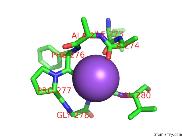
Mono view
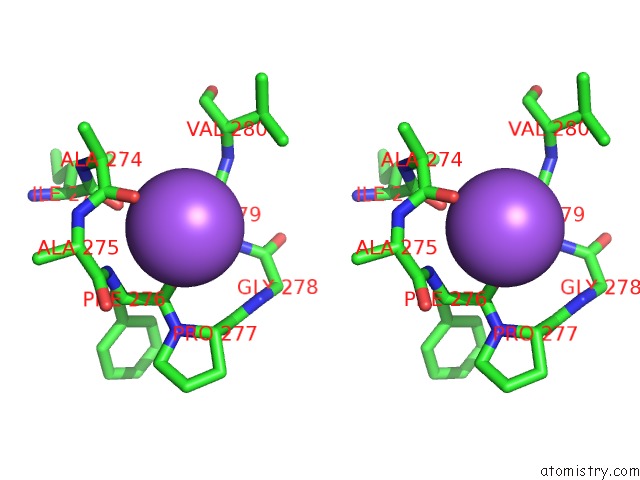
Stereo pair view
Mono view
Stereo pair view
A full contact list of Sodium with other atoms in the Na binding
site number 1 of Crystal Structure of Aspartate Beta-Semialdehide Dehydrogenase From Streptococcus Pneumoniae with 2',5'-Adenosine Diphosphate and D-2- Aminoadipate within 5.0Å range:
|
Reference:
A.G.Pavlovsky,
X.Liu,
C.R.Faehnle,
N.Potente,
R.E.Viola.
Structural Characterization of Inhibitors with Selectivity Against Members of A Homologous Enzyme Family. Chem.Biol.Drug Des. V. 79 128 2012.
ISSN: ISSN 1747-0277
PubMed: 22039970
DOI: 10.1111/J.1747-0285.2011.01267.X
Page generated: Sun Aug 17 16:55:39 2025
ISSN: ISSN 1747-0277
PubMed: 22039970
DOI: 10.1111/J.1747-0285.2011.01267.X
Last articles
Mn in 9LJUMn in 9LJW
Mn in 9LJS
Mn in 9LJR
Mn in 9LJT
Mn in 9LJV
Mg in 9UA2
Mg in 9R96
Mg in 9VM1
Mg in 9P01