Sodium »
PDB 3pkk-3pzs »
3pqh »
Sodium in PDB 3pqh: Crystal Structure of the C-Terminal Fragment of the Bacteriophage PHI92 Membrane-Piercing Protein GP138
Protein crystallography data
The structure of Crystal Structure of the C-Terminal Fragment of the Bacteriophage PHI92 Membrane-Piercing Protein GP138, PDB code: 3pqh
was solved by
C.Browning,
M.Shneider,
P.G.Leiman,
with X-Ray Crystallography technique. A brief refinement statistics is given in the table below:
| Resolution Low / High (Å) | 41.52 / 1.30 |
| Space group | H 3 2 |
| Cell size a, b, c (Å), α, β, γ (°) | 48.080, 48.080, 553.203, 90.00, 90.00, 120.00 |
| R / Rfree (%) | 12.3 / 16 |
Other elements in 3pqh:
The structure of Crystal Structure of the C-Terminal Fragment of the Bacteriophage PHI92 Membrane-Piercing Protein GP138 also contains other interesting chemical elements:
| Iron | (Fe) | 2 atoms |
Sodium Binding Sites:
The binding sites of Sodium atom in the Crystal Structure of the C-Terminal Fragment of the Bacteriophage PHI92 Membrane-Piercing Protein GP138
(pdb code 3pqh). This binding sites where shown within
5.0 Angstroms radius around Sodium atom.
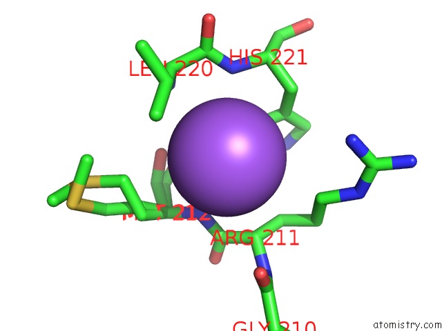
In total 7 binding sites of Sodium where determined in the Crystal Structure of the C-Terminal Fragment of the Bacteriophage PHI92 Membrane-Piercing Protein GP138, PDB code: 3pqh:
Jump to Sodium binding site number: 1; 2; 3; 4; 5; 6; 7;
In total 7 binding sites of Sodium where determined in the Crystal Structure of the C-Terminal Fragment of the Bacteriophage PHI92 Membrane-Piercing Protein GP138, PDB code: 3pqh:
Jump to Sodium binding site number: 1; 2; 3; 4; 5; 6; 7;
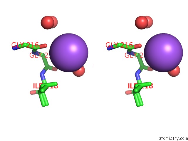
Sodium binding site 1 out of 7 in 3pqh
Go back to
Sodium binding site 1 out
of 7 in the Crystal Structure of the C-Terminal Fragment of the Bacteriophage PHI92 Membrane-Piercing Protein GP138

Mono view
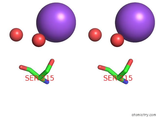
Stereo pair view
Mono view
Stereo pair view
A full contact list of Sodium with other atoms in the Na binding
site number 1 of Crystal Structure of the C-Terminal Fragment of the Bacteriophage PHI92 Membrane-Piercing Protein GP138 within 5.0Å range:
|
Sodium binding site 2 out of 7 in 3pqh
Go back to
Sodium binding site 2 out
of 7 in the Crystal Structure of the C-Terminal Fragment of the Bacteriophage PHI92 Membrane-Piercing Protein GP138
Mono view
Stereo pair view
Mono view
Stereo pair view
A full contact list of Sodium with other atoms in the Na binding
site number 2 of Crystal Structure of the C-Terminal Fragment of the Bacteriophage PHI92 Membrane-Piercing Protein GP138 within 5.0Å range:
|
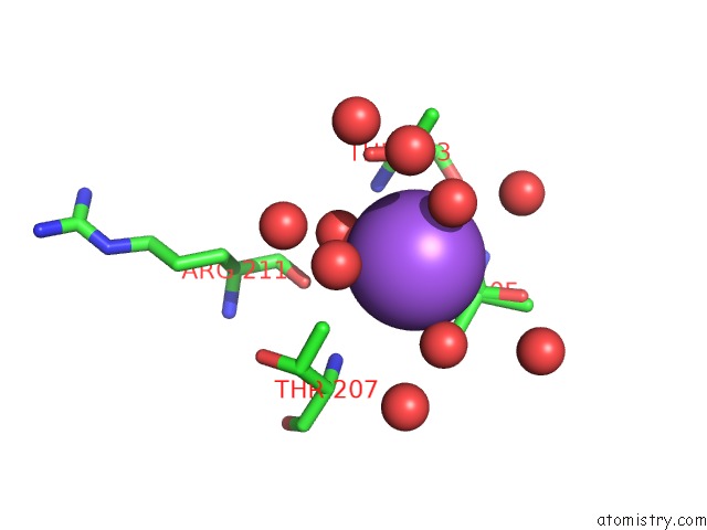
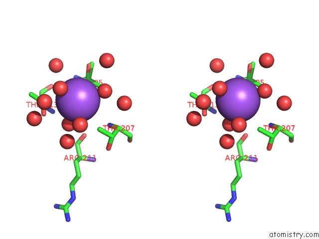
Sodium binding site 3 out of 7 in 3pqh
Go back to
Sodium binding site 3 out
of 7 in the Crystal Structure of the C-Terminal Fragment of the Bacteriophage PHI92 Membrane-Piercing Protein GP138
Mono view

Stereo pair view
Mono view
Stereo pair view
A full contact list of Sodium with other atoms in the Na binding
site number 3 of Crystal Structure of the C-Terminal Fragment of the Bacteriophage PHI92 Membrane-Piercing Protein GP138 within 5.0Å range:
|
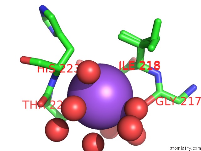
Sodium binding site 4 out of 7 in 3pqh
Go back to
Sodium binding site 4 out
of 7 in the Crystal Structure of the C-Terminal Fragment of the Bacteriophage PHI92 Membrane-Piercing Protein GP138
Mono view

Stereo pair view
Mono view
Stereo pair view
A full contact list of Sodium with other atoms in the Na binding
site number 4 of Crystal Structure of the C-Terminal Fragment of the Bacteriophage PHI92 Membrane-Piercing Protein GP138 within 5.0Å range:
|
Sodium binding site 5 out of 7 in 3pqh
Go back to
Sodium binding site 5 out
of 7 in the Crystal Structure of the C-Terminal Fragment of the Bacteriophage PHI92 Membrane-Piercing Protein GP138

Mono view
Stereo pair view
Mono view
Stereo pair view
A full contact list of Sodium with other atoms in the Na binding
site number 5 of Crystal Structure of the C-Terminal Fragment of the Bacteriophage PHI92 Membrane-Piercing Protein GP138 within 5.0Å range:
|
Sodium binding site 6 out of 7 in 3pqh
Go back to
Sodium binding site 6 out
of 7 in the Crystal Structure of the C-Terminal Fragment of the Bacteriophage PHI92 Membrane-Piercing Protein GP138
Mono view

Stereo pair view
Mono view
Stereo pair view
A full contact list of Sodium with other atoms in the Na binding
site number 6 of Crystal Structure of the C-Terminal Fragment of the Bacteriophage PHI92 Membrane-Piercing Protein GP138 within 5.0Å range:
|
Sodium binding site 7 out of 7 in 3pqh
Go back to
Sodium binding site 7 out
of 7 in the Crystal Structure of the C-Terminal Fragment of the Bacteriophage PHI92 Membrane-Piercing Protein GP138
Mono view

Stereo pair view
Mono view
Stereo pair view
| A full contact list of Sodium with other atoms in the Na binding site number 7 of Crystal Structure of the C-Terminal Fragment of the Bacteriophage PHI92 Membrane-Piercing Protein GP138 within 5.0Å range: |
Reference:
C.Browning,
M.M.Shneider,
V.D.Bowman,
D.Schwarzer,
P.G.Leiman.
Phage Pierces the Host Cell Membrane with the Iron-Loaded Spike. Structure V. 20 326 2012.
ISSN: ISSN 0969-2126
PubMed: 22325780
DOI: 10.1016/J.STR.2011.12.009
Page generated: Sun Aug 17 16:54:37 2025
ISSN: ISSN 0969-2126
PubMed: 22325780
DOI: 10.1016/J.STR.2011.12.009
Last articles
Mn in 9LJUMn in 9LJW
Mn in 9LJS
Mn in 9LJR
Mn in 9LJT
Mn in 9LJV
Mg in 9UA2
Mg in 9R96
Mg in 9VM1
Mg in 9P01