Sodium »
PDB 3ma9-3mo9 »
3mjf »
Sodium in PDB 3mjf: Phosphoribosylamine-Glycine Ligase From Yersinia Pestis
Enzymatic activity of Phosphoribosylamine-Glycine Ligase From Yersinia Pestis
All present enzymatic activity of Phosphoribosylamine-Glycine Ligase From Yersinia Pestis:
6.3.4.13;
6.3.4.13;
Protein crystallography data
The structure of Phosphoribosylamine-Glycine Ligase From Yersinia Pestis, PDB code: 3mjf
was solved by
J.Osipiuk,
M.Zhou,
L.Papazisi,
W.F.Anderson,
A.Joachimiak,
Center Forstructural Genomics Of Infectious Diseases (Csgid),
with X-Ray Crystallography technique. A brief refinement statistics is given in the table below:
| Resolution Low / High (Å) | 30.80 / 1.47 |
| Space group | P 21 21 21 |
| Cell size a, b, c (Å), α, β, γ (°) | 68.637, 69.151, 95.166, 90.00, 90.00, 90.00 |
| R / Rfree (%) | 13.5 / 17.4 |
Sodium Binding Sites:
The binding sites of Sodium atom in the Phosphoribosylamine-Glycine Ligase From Yersinia Pestis
(pdb code 3mjf). This binding sites where shown within
5.0 Angstroms radius around Sodium atom.
In total only one binding site of Sodium was determined in the Phosphoribosylamine-Glycine Ligase From Yersinia Pestis, PDB code: 3mjf:
In total only one binding site of Sodium was determined in the Phosphoribosylamine-Glycine Ligase From Yersinia Pestis, PDB code: 3mjf:
Sodium binding site 1 out of 1 in 3mjf
Go back to
Sodium binding site 1 out
of 1 in the Phosphoribosylamine-Glycine Ligase From Yersinia Pestis
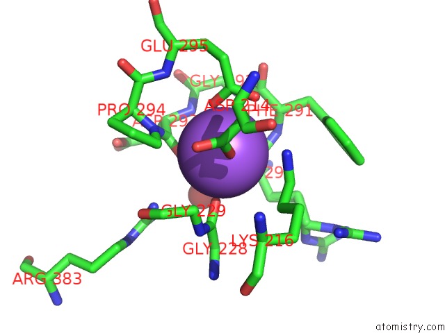
Mono view
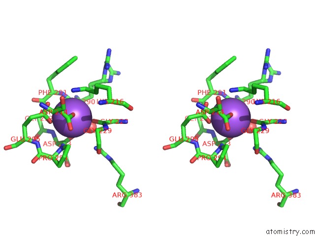
Stereo pair view
Mono view
Stereo pair view
A full contact list of Sodium with other atoms in the Na binding
site number 1 of Phosphoribosylamine-Glycine Ligase From Yersinia Pestis within 5.0Å range:
|
Reference:
J.Osipiuk,
M.Zhou,
L.Papazisi,
W.F.Anderson,
A.Joachimiak.
X-Ray Crystal Structure of Phosphoribosylamine-Glycine Ligase From Yersinia Pestis. To Be Published.
Page generated: Mon Oct 7 11:31:03 2024
Last articles
I in 6YR6I in 6YRB
I in 6YT2
I in 6YGD
I in 6YGC
I in 6YGA
I in 6YGB
I in 6Y54
I in 6XYB
I in 6XYU