Sodium »
PDB 3k9g-3la1 »
3knt »
Sodium in PDB 3knt: Crystal Structure of Methanocaldococcus Jannaschii 8-Oxoguanine Glycosylase/Lyase in Complex with 15MER Dna Containing 8-Oxoguanine
Enzymatic activity of Crystal Structure of Methanocaldococcus Jannaschii 8-Oxoguanine Glycosylase/Lyase in Complex with 15MER Dna Containing 8-Oxoguanine
All present enzymatic activity of Crystal Structure of Methanocaldococcus Jannaschii 8-Oxoguanine Glycosylase/Lyase in Complex with 15MER Dna Containing 8-Oxoguanine:
4.2.99.18;
4.2.99.18;
Protein crystallography data
The structure of Crystal Structure of Methanocaldococcus Jannaschii 8-Oxoguanine Glycosylase/Lyase in Complex with 15MER Dna Containing 8-Oxoguanine, PDB code: 3knt
was solved by
F.Faucher,
S.Doublie,
with X-Ray Crystallography technique. A brief refinement statistics is given in the table below:
| Resolution Low / High (Å) | 19.80 / 2.70 |
| Space group | P 1 21 1 |
| Cell size a, b, c (Å), α, β, γ (°) | 54.800, 150.030, 90.270, 90.00, 107.53, 90.00 |
| R / Rfree (%) | 18.8 / 22.5 |
Sodium Binding Sites:
The binding sites of Sodium atom in the Crystal Structure of Methanocaldococcus Jannaschii 8-Oxoguanine Glycosylase/Lyase in Complex with 15MER Dna Containing 8-Oxoguanine
(pdb code 3knt). This binding sites where shown within
5.0 Angstroms radius around Sodium atom.
In total 4 binding sites of Sodium where determined in the Crystal Structure of Methanocaldococcus Jannaschii 8-Oxoguanine Glycosylase/Lyase in Complex with 15MER Dna Containing 8-Oxoguanine, PDB code: 3knt:
Jump to Sodium binding site number: 1; 2; 3; 4;
In total 4 binding sites of Sodium where determined in the Crystal Structure of Methanocaldococcus Jannaschii 8-Oxoguanine Glycosylase/Lyase in Complex with 15MER Dna Containing 8-Oxoguanine, PDB code: 3knt:
Jump to Sodium binding site number: 1; 2; 3; 4;
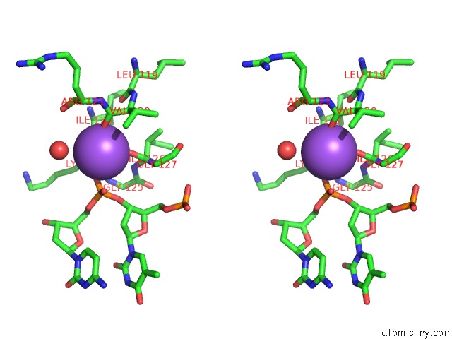
Sodium binding site 1 out of 4 in 3knt
Go back to
Sodium binding site 1 out
of 4 in the Crystal Structure of Methanocaldococcus Jannaschii 8-Oxoguanine Glycosylase/Lyase in Complex with 15MER Dna Containing 8-Oxoguanine
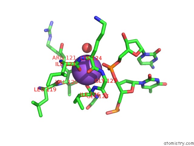
Mono view

Stereo pair view
Mono view
Stereo pair view
A full contact list of Sodium with other atoms in the Na binding
site number 1 of Crystal Structure of Methanocaldococcus Jannaschii 8-Oxoguanine Glycosylase/Lyase in Complex with 15MER Dna Containing 8-Oxoguanine within 5.0Å range:
|
Sodium binding site 2 out of 4 in 3knt
Go back to
Sodium binding site 2 out
of 4 in the Crystal Structure of Methanocaldococcus Jannaschii 8-Oxoguanine Glycosylase/Lyase in Complex with 15MER Dna Containing 8-Oxoguanine
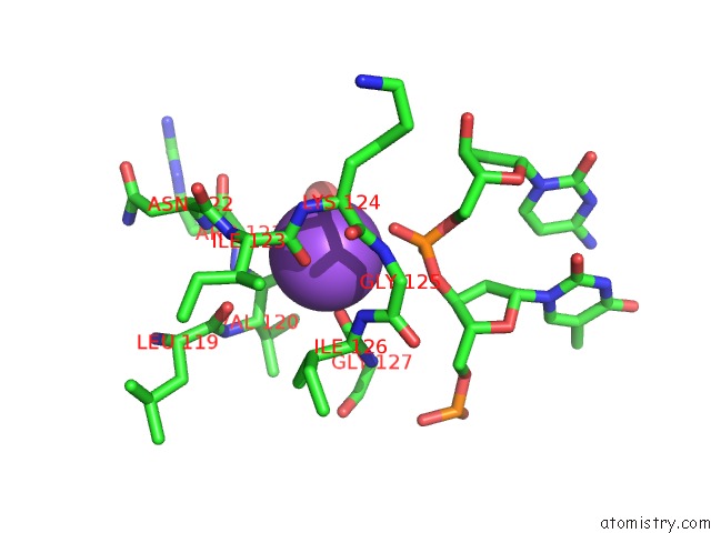
Mono view
Stereo pair view
Mono view
Stereo pair view
A full contact list of Sodium with other atoms in the Na binding
site number 2 of Crystal Structure of Methanocaldococcus Jannaschii 8-Oxoguanine Glycosylase/Lyase in Complex with 15MER Dna Containing 8-Oxoguanine within 5.0Å range:
|
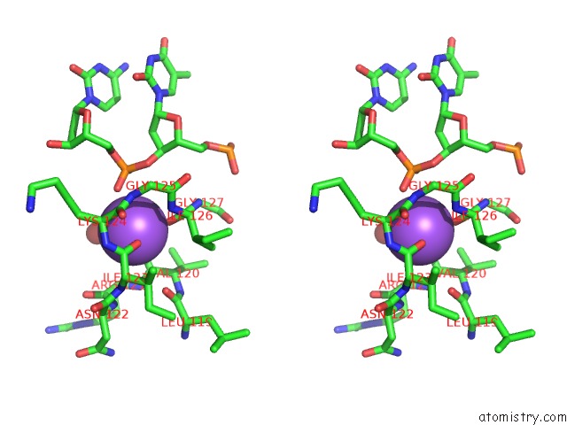
Sodium binding site 3 out of 4 in 3knt
Go back to
Sodium binding site 3 out
of 4 in the Crystal Structure of Methanocaldococcus Jannaschii 8-Oxoguanine Glycosylase/Lyase in Complex with 15MER Dna Containing 8-Oxoguanine
Mono view
Stereo pair view
Mono view
Stereo pair view
A full contact list of Sodium with other atoms in the Na binding
site number 3 of Crystal Structure of Methanocaldococcus Jannaschii 8-Oxoguanine Glycosylase/Lyase in Complex with 15MER Dna Containing 8-Oxoguanine within 5.0Å range:
|
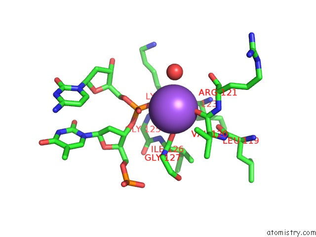
Sodium binding site 4 out of 4 in 3knt
Go back to
Sodium binding site 4 out
of 4 in the Crystal Structure of Methanocaldococcus Jannaschii 8-Oxoguanine Glycosylase/Lyase in Complex with 15MER Dna Containing 8-Oxoguanine

Mono view

Stereo pair view
Mono view
Stereo pair view
A full contact list of Sodium with other atoms in the Na binding
site number 4 of Crystal Structure of Methanocaldococcus Jannaschii 8-Oxoguanine Glycosylase/Lyase in Complex with 15MER Dna Containing 8-Oxoguanine within 5.0Å range:
|
Reference:
F.Faucher,
S.S.Wallace,
S.Doublie.
The C-Terminal Lysine of OGG2 Dna Glycosylases Is A Major Molecular Determinant For Guanine/8-Oxoguanine Distinction. J.Mol.Biol. V. 397 46 2010.
ISSN: ISSN 0022-2836
PubMed: 20083120
DOI: 10.1016/J.JMB.2010.01.024
Page generated: Mon Oct 7 11:13:52 2024
ISSN: ISSN 0022-2836
PubMed: 20083120
DOI: 10.1016/J.JMB.2010.01.024
Last articles
Zn in 9MJ5Zn in 9HNW
Zn in 9G0L
Zn in 9FNE
Zn in 9DZN
Zn in 9E0I
Zn in 9D32
Zn in 9DAK
Zn in 8ZXC
Zn in 8ZUF