Sodium »
PDB 3icf-3ivu »
3ird »
Sodium in PDB 3ird: Structure of Dihydrodipicolinate Synthase From Clostridium Botulinum
Enzymatic activity of Structure of Dihydrodipicolinate Synthase From Clostridium Botulinum
All present enzymatic activity of Structure of Dihydrodipicolinate Synthase From Clostridium Botulinum:
4.2.1.52;
4.2.1.52;
Protein crystallography data
The structure of Structure of Dihydrodipicolinate Synthase From Clostridium Botulinum, PDB code: 3ird
was solved by
R.C.J.Dobson,
S.Atkinson,
M.A.Perugini,
with X-Ray Crystallography technique. A brief refinement statistics is given in the table below:
| Resolution Low / High (Å) | 34.20 / 2.23 |
| Space group | P 42 21 2 |
| Cell size a, b, c (Å), α, β, γ (°) | 92.810, 92.810, 60.350, 90.00, 90.00, 90.00 |
| R / Rfree (%) | 13.5 / 20.9 |
Other elements in 3ird:
The structure of Structure of Dihydrodipicolinate Synthase From Clostridium Botulinum also contains other interesting chemical elements:
| Chlorine | (Cl) | 2 atoms |
Sodium Binding Sites:
The binding sites of Sodium atom in the Structure of Dihydrodipicolinate Synthase From Clostridium Botulinum
(pdb code 3ird). This binding sites where shown within
5.0 Angstroms radius around Sodium atom.
In total only one binding site of Sodium was determined in the Structure of Dihydrodipicolinate Synthase From Clostridium Botulinum, PDB code: 3ird:
In total only one binding site of Sodium was determined in the Structure of Dihydrodipicolinate Synthase From Clostridium Botulinum, PDB code: 3ird:
Sodium binding site 1 out of 1 in 3ird
Go back to
Sodium binding site 1 out
of 1 in the Structure of Dihydrodipicolinate Synthase From Clostridium Botulinum
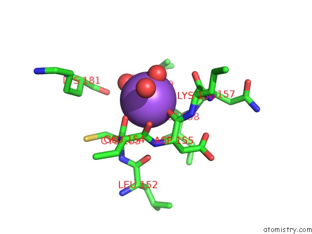
Mono view
Stereo pair view
Mono view
Stereo pair view
A full contact list of Sodium with other atoms in the Na binding
site number 1 of Structure of Dihydrodipicolinate Synthase From Clostridium Botulinum within 5.0Å range:
|
Reference:
S.Atkinson,
R.C.J.Dobson,
M.A.Perugini.
Structure of Cbot-Dhdps To Be Published.
Page generated: Mon Oct 7 11:01:31 2024
Last articles
Mn in 5WEYMn in 5WEI
Mn in 5WEB
Mn in 5WEF
Mn in 5WDY
Mn in 5WE8
Mn in 5WE7
Mn in 5WE9
Mn in 5WDW
Mn in 5WCX