Sodium »
PDB 3gdx-3h09 »
3geo »
Sodium in PDB 3geo: Sulfite Reductase Hemoprotein Nitrite Complex
Enzymatic activity of Sulfite Reductase Hemoprotein Nitrite Complex
All present enzymatic activity of Sulfite Reductase Hemoprotein Nitrite Complex:
1.8.1.2;
1.8.1.2;
Protein crystallography data
The structure of Sulfite Reductase Hemoprotein Nitrite Complex, PDB code: 3geo
was solved by
B.R.Crane,
E.D.Getzoff,
with X-Ray Crystallography technique. A brief refinement statistics is given in the table below:
| Resolution Low / High (Å) | 10.00 / 2.10 |
| Space group | P 21 21 21 |
| Cell size a, b, c (Å), α, β, γ (°) | 69.800, 77.400, 87.800, 90.00, 90.00, 90.00 |
| R / Rfree (%) | 18.5 / n/a |
Other elements in 3geo:
The structure of Sulfite Reductase Hemoprotein Nitrite Complex also contains other interesting chemical elements:
| Iron | (Fe) | 5 atoms |
Sodium Binding Sites:
The binding sites of Sodium atom in the Sulfite Reductase Hemoprotein Nitrite Complex
(pdb code 3geo). This binding sites where shown within
5.0 Angstroms radius around Sodium atom.
In total only one binding site of Sodium was determined in the Sulfite Reductase Hemoprotein Nitrite Complex, PDB code: 3geo:
In total only one binding site of Sodium was determined in the Sulfite Reductase Hemoprotein Nitrite Complex, PDB code: 3geo:
Sodium binding site 1 out of 1 in 3geo
Go back to
Sodium binding site 1 out
of 1 in the Sulfite Reductase Hemoprotein Nitrite Complex
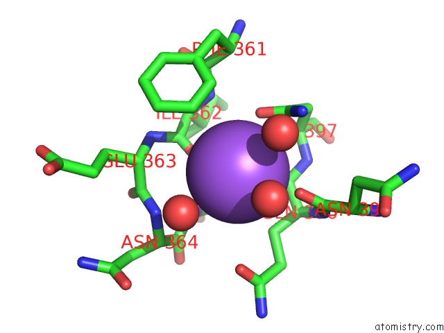
Mono view
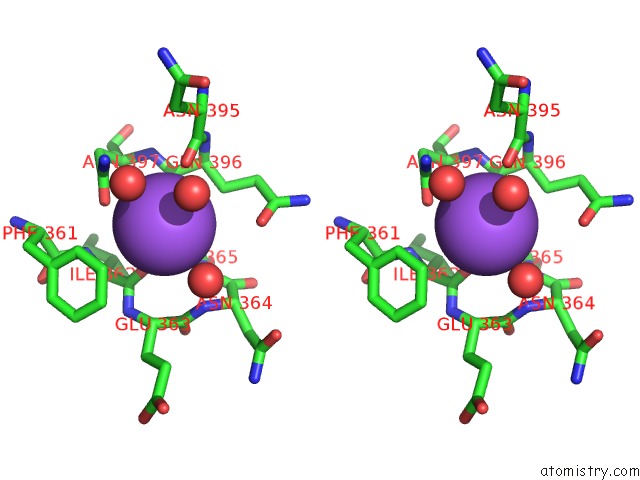
Stereo pair view
Mono view
Stereo pair view
A full contact list of Sodium with other atoms in the Na binding
site number 1 of Sulfite Reductase Hemoprotein Nitrite Complex within 5.0Å range:
|
Reference:
B.R.Crane,
L.M.Siegel,
E.D.Getzoff.
Probing the Catalytic Mechanism of Sulfite Reductase By X-Ray Crystallography: Structures of the Escherichia Coli Hemoprotein in Complex with Substrates, Inhibitors, Intermediates, and Products. Biochemistry V. 36 12120 1997.
ISSN: ISSN 0006-2960
PubMed: 9315849
DOI: 10.1021/BI971066I
Page generated: Mon Oct 7 10:15:08 2024
ISSN: ISSN 0006-2960
PubMed: 9315849
DOI: 10.1021/BI971066I
Last articles
Mg in 2UU9Mg in 2UU7
Mg in 2UAG
Mg in 2UKD
Mg in 2SHK
Mg in 2TPS
Mg in 2TRT
Mg in 2TRA
Mg in 2RMK
Mg in 2RUS