Sodium »
PDB 3e40-3ept »
3eoj »
Sodium in PDB 3eoj: Fmo Protein From Prosthecochloris Aestuarii 2K at 1.3A Resolution
Protein crystallography data
The structure of Fmo Protein From Prosthecochloris Aestuarii 2K at 1.3A Resolution, PDB code: 3eoj
was solved by
D.E.Tronrud,
J.Wen,
L.Gay,
R.E.Blankenship,
with X-Ray Crystallography technique. A brief refinement statistics is given in the table below:
| Resolution Low / High (Å) | 50.00 / 1.30 |
| Space group | P 63 |
| Cell size a, b, c (Å), α, β, γ (°) | 111.242, 111.242, 98.198, 90.00, 90.00, 120.00 |
| R / Rfree (%) | 13.5 / 16.1 |
Other elements in 3eoj:
The structure of Fmo Protein From Prosthecochloris Aestuarii 2K at 1.3A Resolution also contains other interesting chemical elements:
| Magnesium | (Mg) | 8 atoms |
Sodium Binding Sites:
The binding sites of Sodium atom in the Fmo Protein From Prosthecochloris Aestuarii 2K at 1.3A Resolution
(pdb code 3eoj). This binding sites where shown within
5.0 Angstroms radius around Sodium atom.
In total only one binding site of Sodium was determined in the Fmo Protein From Prosthecochloris Aestuarii 2K at 1.3A Resolution, PDB code: 3eoj:
In total only one binding site of Sodium was determined in the Fmo Protein From Prosthecochloris Aestuarii 2K at 1.3A Resolution, PDB code: 3eoj:
Sodium binding site 1 out of 1 in 3eoj
Go back to
Sodium binding site 1 out
of 1 in the Fmo Protein From Prosthecochloris Aestuarii 2K at 1.3A Resolution
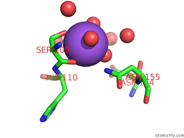
Mono view
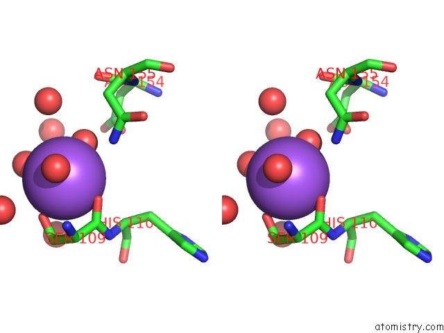
Stereo pair view
Mono view
Stereo pair view
A full contact list of Sodium with other atoms in the Na binding
site number 1 of Fmo Protein From Prosthecochloris Aestuarii 2K at 1.3A Resolution within 5.0Å range:
|
Reference:
D.E.Tronrud,
J.Wen,
L.Gay,
R.E.Blankenship.
The Structural Basis For the Difference in Absorbance Spectra For the Fmo Antenna Protein From Various Green Sulfur Bacteria. Photosynth.Res. V. 100 79 2009.
ISSN: ISSN 0166-8595
PubMed: 19437128
DOI: 10.1007/S11120-009-9430-6
Page generated: Sun Aug 17 14:42:50 2025
ISSN: ISSN 0166-8595
PubMed: 19437128
DOI: 10.1007/S11120-009-9430-6
Last articles
Mn in 9LJUMn in 9LJW
Mn in 9LJS
Mn in 9LJR
Mn in 9LJT
Mn in 9LJV
Mg in 9UA2
Mg in 9R96
Mg in 9VM1
Mg in 9P01