Sodium »
PDB 3e40-3ept »
3eat »
Sodium in PDB 3eat: Crystal Structure of the Pvcb (PA2255) Protein From Pseudomonas Aeruginosa
Protein crystallography data
The structure of Crystal Structure of the Pvcb (PA2255) Protein From Pseudomonas Aeruginosa, PDB code: 3eat
was solved by
A.M.Gulick,
E.J.Drake,
with X-Ray Crystallography technique. A brief refinement statistics is given in the table below:
| Resolution Low / High (Å) | 30.00 / 2.50 |
| Space group | P 63 2 2 |
| Cell size a, b, c (Å), α, β, γ (°) | 125.350, 125.350, 107.110, 90.00, 90.00, 120.00 |
| R / Rfree (%) | 18.7 / 22.5 |
Sodium Binding Sites:
The binding sites of Sodium atom in the Crystal Structure of the Pvcb (PA2255) Protein From Pseudomonas Aeruginosa
(pdb code 3eat). This binding sites where shown within
5.0 Angstroms radius around Sodium atom.
In total only one binding site of Sodium was determined in the Crystal Structure of the Pvcb (PA2255) Protein From Pseudomonas Aeruginosa, PDB code: 3eat:
In total only one binding site of Sodium was determined in the Crystal Structure of the Pvcb (PA2255) Protein From Pseudomonas Aeruginosa, PDB code: 3eat:
Sodium binding site 1 out of 1 in 3eat
Go back to
Sodium binding site 1 out
of 1 in the Crystal Structure of the Pvcb (PA2255) Protein From Pseudomonas Aeruginosa
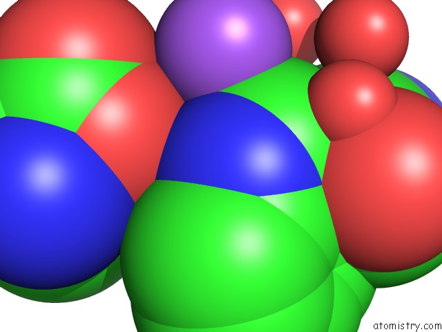
Mono view
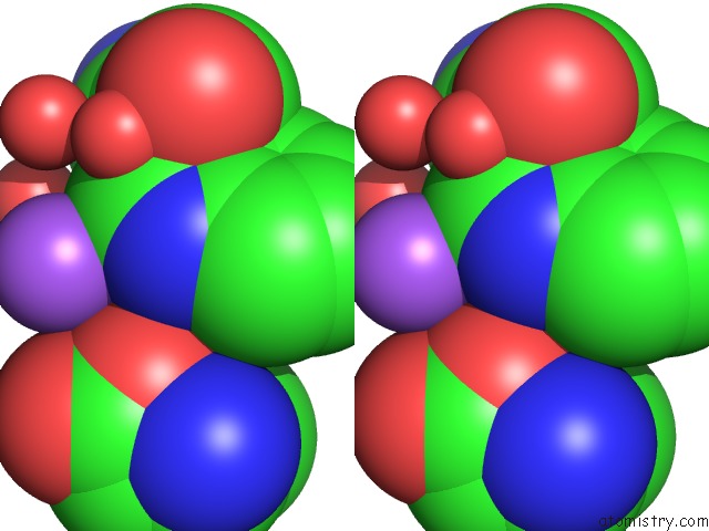
Stereo pair view
Mono view
Stereo pair view
A full contact list of Sodium with other atoms in the Na binding
site number 1 of Crystal Structure of the Pvcb (PA2255) Protein From Pseudomonas Aeruginosa within 5.0Å range:
|
Reference:
E.J.Drake,
A.M.Gulick.
Three-Dimensional Structures of Pseudomonas Aeruginosa Pvca and Pvcb, Two Proteins Involved in the Synthesis of 2-Isocyano-6,7-Dihydroxycoumarin. J.Mol.Biol. V. 384 193 2008.
ISSN: ISSN 0022-2836
PubMed: 18824174
DOI: 10.1016/J.JMB.2008.09.027
Page generated: Sun Aug 17 14:41:09 2025
ISSN: ISSN 0022-2836
PubMed: 18824174
DOI: 10.1016/J.JMB.2008.09.027
Last articles
Mn in 9LJUMn in 9LJW
Mn in 9LJS
Mn in 9LJR
Mn in 9LJT
Mn in 9LJV
Mg in 9UA2
Mg in 9R96
Mg in 9VM1
Mg in 9P01