Sodium »
PDB 3cq8-3dfh »
3ddr »
Sodium in PDB 3ddr: Structure of the Serratia Marcescens Hemophore Receptor Hasr-ILE671GLY Mutant in Complex with Its Hemophore Hasa and Heme
Protein crystallography data
The structure of Structure of the Serratia Marcescens Hemophore Receptor Hasr-ILE671GLY Mutant in Complex with Its Hemophore Hasa and Heme, PDB code: 3ddr
was solved by
S.Krieg,
K.Diederichs,
with X-Ray Crystallography technique. A brief refinement statistics is given in the table below:
| Resolution Low / High (Å) | 39.25 / 2.80 |
| Space group | F 2 2 2 |
| Cell size a, b, c (Å), α, β, γ (°) | 157.410, 162.720, 597.010, 90.00, 90.00, 90.00 |
| R / Rfree (%) | 22.6 / 26.2 |
Other elements in 3ddr:
The structure of Structure of the Serratia Marcescens Hemophore Receptor Hasr-ILE671GLY Mutant in Complex with Its Hemophore Hasa and Heme also contains other interesting chemical elements:
| Iron | (Fe) | 2 atoms |
Sodium Binding Sites:
The binding sites of Sodium atom in the Structure of the Serratia Marcescens Hemophore Receptor Hasr-ILE671GLY Mutant in Complex with Its Hemophore Hasa and Heme
(pdb code 3ddr). This binding sites where shown within
5.0 Angstroms radius around Sodium atom.
In total 2 binding sites of Sodium where determined in the Structure of the Serratia Marcescens Hemophore Receptor Hasr-ILE671GLY Mutant in Complex with Its Hemophore Hasa and Heme, PDB code: 3ddr:
Jump to Sodium binding site number: 1; 2;
In total 2 binding sites of Sodium where determined in the Structure of the Serratia Marcescens Hemophore Receptor Hasr-ILE671GLY Mutant in Complex with Its Hemophore Hasa and Heme, PDB code: 3ddr:
Jump to Sodium binding site number: 1; 2;
Sodium binding site 1 out of 2 in 3ddr
Go back to
Sodium binding site 1 out
of 2 in the Structure of the Serratia Marcescens Hemophore Receptor Hasr-ILE671GLY Mutant in Complex with Its Hemophore Hasa and Heme
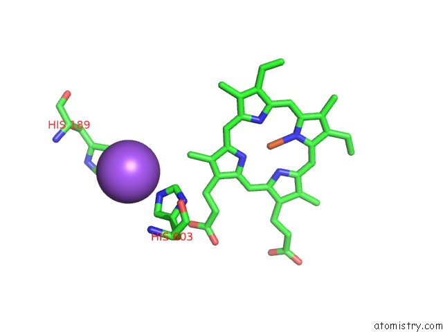
Mono view
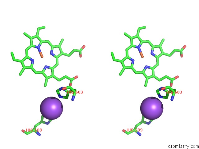
Stereo pair view
Mono view
Stereo pair view
A full contact list of Sodium with other atoms in the Na binding
site number 1 of Structure of the Serratia Marcescens Hemophore Receptor Hasr-ILE671GLY Mutant in Complex with Its Hemophore Hasa and Heme within 5.0Å range:
|
Sodium binding site 2 out of 2 in 3ddr
Go back to
Sodium binding site 2 out
of 2 in the Structure of the Serratia Marcescens Hemophore Receptor Hasr-ILE671GLY Mutant in Complex with Its Hemophore Hasa and Heme
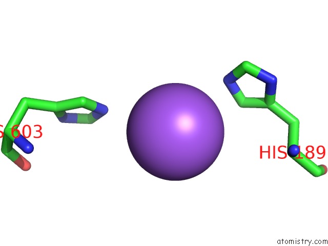
Mono view
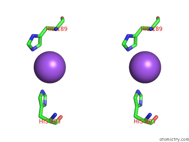
Stereo pair view
Mono view
Stereo pair view
A full contact list of Sodium with other atoms in the Na binding
site number 2 of Structure of the Serratia Marcescens Hemophore Receptor Hasr-ILE671GLY Mutant in Complex with Its Hemophore Hasa and Heme within 5.0Å range:
|
Reference:
S.Krieg,
F.Huche,
K.Diederichs,
N.Izadi-Pruneyre,
A.Lecroisey,
C.Wandersman,
P.Delepelaire,
W.Welte.
Heme Uptake Across the Outer Membrane As Revealed By Crystal Structures of the Receptor-Hemophore Complex. Proc.Natl.Acad.Sci.Usa V. 106 1045 2009.
ISSN: ISSN 0027-8424
PubMed: 19144921
DOI: 10.1073/PNAS.0809406106
Page generated: Mon Oct 7 08:12:29 2024
ISSN: ISSN 0027-8424
PubMed: 19144921
DOI: 10.1073/PNAS.0809406106
Last articles
K in 3P1EK in 3P1D
K in 3OYZ
K in 3OYX
K in 3P1C
K in 3OYT
K in 3P10
K in 3P0Z
K in 3OTO
K in 3OUF