Sodium »
PDB 3c7o-3cpw »
3c7o »
Sodium in PDB 3c7o: Crystal Structure of A Glycoside Hydrolase Family 43 Arabinoxylan Arabinofuranohydrolase From Bacillus Subtilis in Complex with Cellotetraose.
Enzymatic activity of Crystal Structure of A Glycoside Hydrolase Family 43 Arabinoxylan Arabinofuranohydrolase From Bacillus Subtilis in Complex with Cellotetraose.
All present enzymatic activity of Crystal Structure of A Glycoside Hydrolase Family 43 Arabinoxylan Arabinofuranohydrolase From Bacillus Subtilis in Complex with Cellotetraose.:
3.2.1.55;
3.2.1.55;
Protein crystallography data
The structure of Crystal Structure of A Glycoside Hydrolase Family 43 Arabinoxylan Arabinofuranohydrolase From Bacillus Subtilis in Complex with Cellotetraose., PDB code: 3c7o
was solved by
E.Vandermarliere,
T.M.Bourgois,
M.D.Winn,
S.Van Campenhout,
G.Volckaert,
S.V.Strelkov,
J.A.Delcour,
A.Rabijns,
C.M.Courtin,
with X-Ray Crystallography technique. A brief refinement statistics is given in the table below:
| Resolution Low / High (Å) | 29.59 / 1.80 |
| Space group | P 21 21 21 |
| Cell size a, b, c (Å), α, β, γ (°) | 67.990, 73.080, 105.690, 90.00, 90.00, 90.00 |
| R / Rfree (%) | 15.1 / 17.5 |
Other elements in 3c7o:
The structure of Crystal Structure of A Glycoside Hydrolase Family 43 Arabinoxylan Arabinofuranohydrolase From Bacillus Subtilis in Complex with Cellotetraose. also contains other interesting chemical elements:
| Calcium | (Ca) | 1 atom |
Sodium Binding Sites:
The binding sites of Sodium atom in the Crystal Structure of A Glycoside Hydrolase Family 43 Arabinoxylan Arabinofuranohydrolase From Bacillus Subtilis in Complex with Cellotetraose.
(pdb code 3c7o). This binding sites where shown within
5.0 Angstroms radius around Sodium atom.
In total 2 binding sites of Sodium where determined in the Crystal Structure of A Glycoside Hydrolase Family 43 Arabinoxylan Arabinofuranohydrolase From Bacillus Subtilis in Complex with Cellotetraose., PDB code: 3c7o:
Jump to Sodium binding site number: 1; 2;
In total 2 binding sites of Sodium where determined in the Crystal Structure of A Glycoside Hydrolase Family 43 Arabinoxylan Arabinofuranohydrolase From Bacillus Subtilis in Complex with Cellotetraose., PDB code: 3c7o:
Jump to Sodium binding site number: 1; 2;
Sodium binding site 1 out of 2 in 3c7o
Go back to
Sodium binding site 1 out
of 2 in the Crystal Structure of A Glycoside Hydrolase Family 43 Arabinoxylan Arabinofuranohydrolase From Bacillus Subtilis in Complex with Cellotetraose.

Mono view
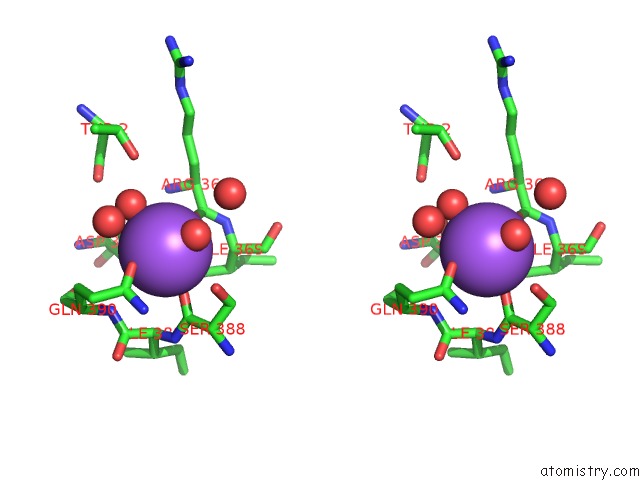
Stereo pair view
Mono view
Stereo pair view
A full contact list of Sodium with other atoms in the Na binding
site number 1 of Crystal Structure of A Glycoside Hydrolase Family 43 Arabinoxylan Arabinofuranohydrolase From Bacillus Subtilis in Complex with Cellotetraose. within 5.0Å range:
|
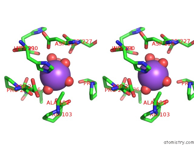
Sodium binding site 2 out of 2 in 3c7o
Go back to
Sodium binding site 2 out
of 2 in the Crystal Structure of A Glycoside Hydrolase Family 43 Arabinoxylan Arabinofuranohydrolase From Bacillus Subtilis in Complex with Cellotetraose.
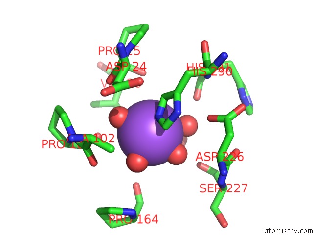
Mono view
Stereo pair view
Mono view
Stereo pair view
A full contact list of Sodium with other atoms in the Na binding
site number 2 of Crystal Structure of A Glycoside Hydrolase Family 43 Arabinoxylan Arabinofuranohydrolase From Bacillus Subtilis in Complex with Cellotetraose. within 5.0Å range:
|
Reference:
E.Vandermarliere,
T.M.Bourgois,
M.D.Winn,
S.Van Campenhout,
G.Volckaert,
J.A.Delcour,
S.V.Strelkov,
A.Rabijns,
C.M.Courtin.
Structural Analysis of A Glycoside Hydrolase Family 43 Arabinoxylan Arabinofuranohydrolase in Complex with Xylotetraose Reveals A Different Binding Mechanism Compared with Other Members of the Same Family. Biochem.J. V. 418 39 2009.
ISSN: ISSN 0264-6021
PubMed: 18980579
DOI: 10.1042/BJ20081256
Page generated: Sun Aug 17 12:57:20 2025
ISSN: ISSN 0264-6021
PubMed: 18980579
DOI: 10.1042/BJ20081256
Last articles
K in 9NESK in 9PHG
K in 9NEI
K in 9NED
K in 9NEC
K in 9NEG
K in 9CWU
K in 9CVB
K in 9CVA
K in 9COM