Sodium »
PDB 3bjp-3c7h »
3bpx »
Sodium in PDB 3bpx: Crystal Structure of Marr
Protein crystallography data
The structure of Crystal Structure of Marr, PDB code: 3bpx
was solved by
V.Saridakis,
D.Shahinas,
X.Xu,
D.Christendat,
with X-Ray Crystallography technique. A brief refinement statistics is given in the table below:
| Resolution Low / High (Å) | 26.29 / 1.95 |
| Space group | P 21 21 21 |
| Cell size a, b, c (Å), α, β, γ (°) | 40.109, 51.527, 139.243, 90.00, 90.00, 90.00 |
| R / Rfree (%) | 22.2 / 28.4 |
Sodium Binding Sites:
The binding sites of Sodium atom in the Crystal Structure of Marr
(pdb code 3bpx). This binding sites where shown within
5.0 Angstroms radius around Sodium atom.
In total 6 binding sites of Sodium where determined in the Crystal Structure of Marr, PDB code: 3bpx:
Jump to Sodium binding site number: 1; 2; 3; 4; 5; 6;
In total 6 binding sites of Sodium where determined in the Crystal Structure of Marr, PDB code: 3bpx:
Jump to Sodium binding site number: 1; 2; 3; 4; 5; 6;
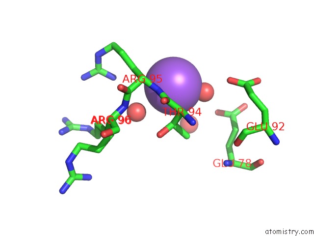
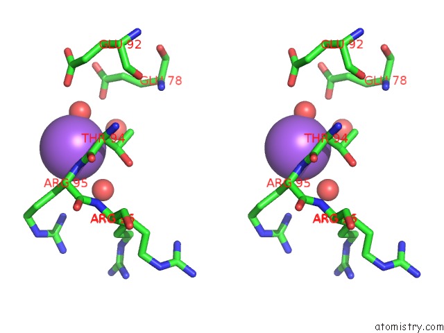
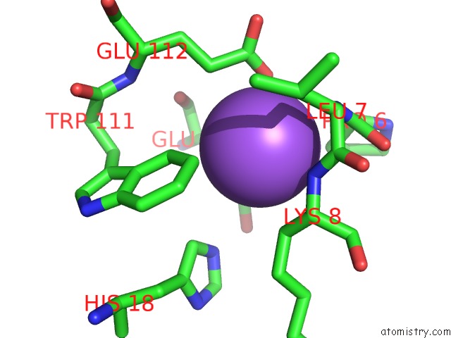
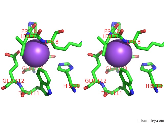
Sodium binding site 1 out of 6 in 3bpx
Go back to
Sodium binding site 1 out
of 6 in the Crystal Structure of Marr
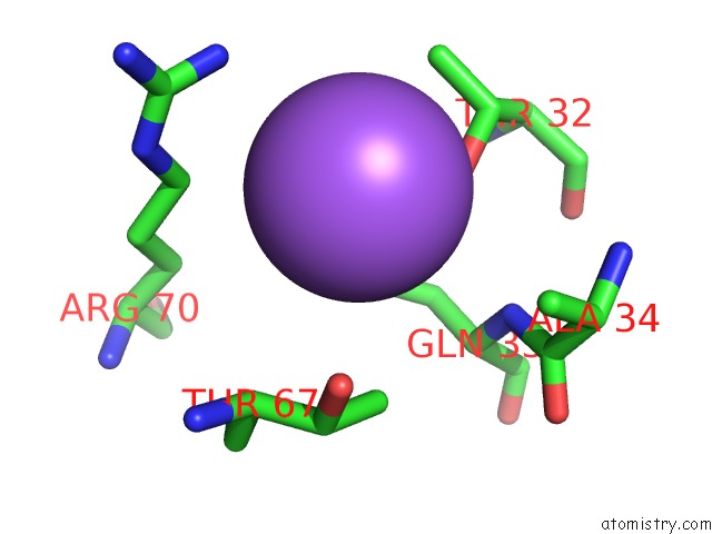
Mono view
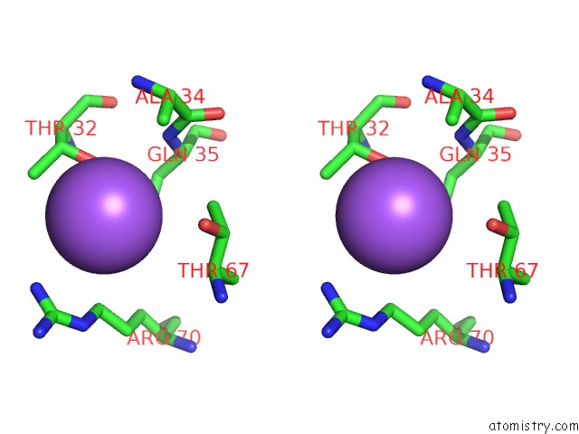
Stereo pair view
Mono view
Stereo pair view
A full contact list of Sodium with other atoms in the Na binding
site number 1 of Crystal Structure of Marr within 5.0Å range:
|
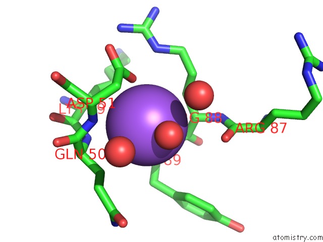
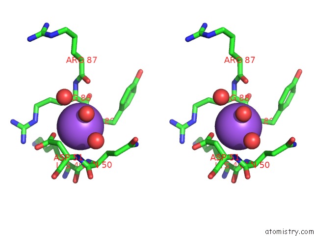
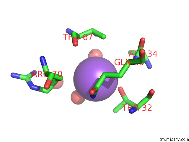
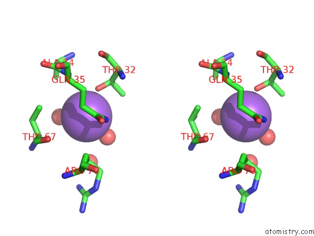
Sodium binding site 2 out of 6 in 3bpx
Go back to
Sodium binding site 2 out
of 6 in the Crystal Structure of Marr
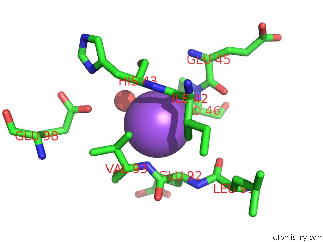
Mono view
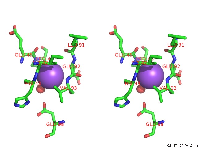
Stereo pair view
Mono view
Stereo pair view
A full contact list of Sodium with other atoms in the Na binding
site number 2 of Crystal Structure of Marr within 5.0Å range:
|
Sodium binding site 3 out of 6 in 3bpx
Go back to
Sodium binding site 3 out
of 6 in the Crystal Structure of Marr
Mono view
Stereo pair view
Mono view
Stereo pair view
A full contact list of Sodium with other atoms in the Na binding
site number 3 of Crystal Structure of Marr within 5.0Å range:
|
Sodium binding site 4 out of 6 in 3bpx
Go back to
Sodium binding site 4 out
of 6 in the Crystal Structure of Marr
Mono view
Stereo pair view
Mono view
Stereo pair view
A full contact list of Sodium with other atoms in the Na binding
site number 4 of Crystal Structure of Marr within 5.0Å range:
|
Sodium binding site 5 out of 6 in 3bpx
Go back to
Sodium binding site 5 out
of 6 in the Crystal Structure of Marr
Mono view
Stereo pair view
Mono view
Stereo pair view
A full contact list of Sodium with other atoms in the Na binding
site number 5 of Crystal Structure of Marr within 5.0Å range:
|
Sodium binding site 6 out of 6 in 3bpx
Go back to
Sodium binding site 6 out
of 6 in the Crystal Structure of Marr
Mono view
Stereo pair view
Mono view
Stereo pair view
A full contact list of Sodium with other atoms in the Na binding
site number 6 of Crystal Structure of Marr within 5.0Å range:
|
Reference:
V.Saridakis,
D.Shahinas,
X.Xu,
D.Christendat.
Structural Insight on the Mechanism of Regulation of the Marr Family of Proteins: High-Resolution Crystal Structure of A Transcriptional Repressor From Methanobacterium Thermoautotrophicum. J.Mol.Biol. V. 377 655 2008.
ISSN: ISSN 0022-2836
PubMed: 18272181
DOI: 10.1016/J.JMB.2008.01.001
Page generated: Sun Aug 17 12:52:37 2025
ISSN: ISSN 0022-2836
PubMed: 18272181
DOI: 10.1016/J.JMB.2008.01.001
Last articles
Mn in 9LJUMn in 9LJW
Mn in 9LJS
Mn in 9LJR
Mn in 9LJT
Mn in 9LJV
Mg in 9UA2
Mg in 9R96
Mg in 9VM1
Mg in 9P01