Sodium »
PDB 2zgx-3a03 »
378d »
Sodium in PDB 378d: Structure of the Side-By-Side Binding of Distamycin to Dna
Protein crystallography data
The structure of Structure of the Side-By-Side Binding of Distamycin to Dna, PDB code: 378d
was solved by
S.N.Mitra,
M.C.Wahl,
M.Sundaralingam,
with X-Ray Crystallography technique. A brief refinement statistics is given in the table below:
| Resolution Low / High (Å) | 8.00 / 2.40 |
| Space group | P 1 21 1 |
| Cell size a, b, c (Å), α, β, γ (°) | 29.550, 42.180, 43.380, 90.00, 96.56, 90.00 |
| R / Rfree (%) | 21 / 28.6 |
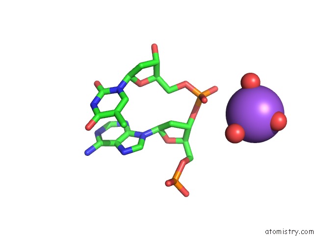
Sodium Binding Sites:
The binding sites of Sodium atom in the Structure of the Side-By-Side Binding of Distamycin to Dna
(pdb code 378d). This binding sites where shown within
5.0 Angstroms radius around Sodium atom.
In total only one binding site of Sodium was determined in the Structure of the Side-By-Side Binding of Distamycin to Dna, PDB code: 378d:
In total only one binding site of Sodium was determined in the Structure of the Side-By-Side Binding of Distamycin to Dna, PDB code: 378d:
Sodium binding site 1 out of 1 in 378d
Go back to
Sodium binding site 1 out
of 1 in the Structure of the Side-By-Side Binding of Distamycin to Dna
Mono view
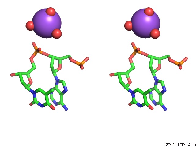
Stereo pair view
Mono view
Stereo pair view
A full contact list of Sodium with other atoms in the Na binding
site number 1 of Structure of the Side-By-Side Binding of Distamycin to Dna within 5.0Å range:
|
Reference:
S.N.Mitra,
M.C.Wahl,
M.Sundaralingam.
Structure of the Side-By-Side Binding of Distamycin to D(Gtatatac)2. Acta Crystallogr.,Sect.D V. 55 602 1999.
ISSN: ISSN 0907-4449
PubMed: 10089456
DOI: 10.1107/S0907444998012475
Page generated: Sun Aug 17 12:36:14 2025
ISSN: ISSN 0907-4449
PubMed: 10089456
DOI: 10.1107/S0907444998012475
Last articles
Mn in 9LJUMn in 9LJW
Mn in 9LJS
Mn in 9LJR
Mn in 9LJT
Mn in 9LJV
Mg in 9UA2
Mg in 9R96
Mg in 9VM1
Mg in 9P01