Sodium »
PDB 2zgx-3a03 »
2zj9 »
Sodium in PDB 2zj9: X-Ray Crystal Structure of Ampc Beta-Lactamase (Ampc(D)) From An Escherichia Coli with A Tripeptide Deletion (GLY286 SER287 ASP288) on the H10 Helix
Enzymatic activity of X-Ray Crystal Structure of Ampc Beta-Lactamase (Ampc(D)) From An Escherichia Coli with A Tripeptide Deletion (GLY286 SER287 ASP288) on the H10 Helix
All present enzymatic activity of X-Ray Crystal Structure of Ampc Beta-Lactamase (Ampc(D)) From An Escherichia Coli with A Tripeptide Deletion (GLY286 SER287 ASP288) on the H10 Helix:
3.5.2.6;
3.5.2.6;
Protein crystallography data
The structure of X-Ray Crystal Structure of Ampc Beta-Lactamase (Ampc(D)) From An Escherichia Coli with A Tripeptide Deletion (GLY286 SER287 ASP288) on the H10 Helix, PDB code: 2zj9
was solved by
Y.Yamaguchi,
G.Sato,
Y.Yamagata,
J.Wachino,
Y.Arakawa,
H.Kurosaki,
with X-Ray Crystallography technique. A brief refinement statistics is given in the table below:
| Resolution Low / High (Å) | 39.16 / 1.70 |
| Space group | P 1 |
| Cell size a, b, c (Å), α, β, γ (°) | 47.065, 47.381, 81.461, 82.62, 80.91, 65.40 |
| R / Rfree (%) | 16.3 / 20.6 |
Sodium Binding Sites:
The binding sites of Sodium atom in the X-Ray Crystal Structure of Ampc Beta-Lactamase (Ampc(D)) From An Escherichia Coli with A Tripeptide Deletion (GLY286 SER287 ASP288) on the H10 Helix
(pdb code 2zj9). This binding sites where shown within
5.0 Angstroms radius around Sodium atom.
In total 2 binding sites of Sodium where determined in the X-Ray Crystal Structure of Ampc Beta-Lactamase (Ampc(D)) From An Escherichia Coli with A Tripeptide Deletion (GLY286 SER287 ASP288) on the H10 Helix, PDB code: 2zj9:
Jump to Sodium binding site number: 1; 2;
In total 2 binding sites of Sodium where determined in the X-Ray Crystal Structure of Ampc Beta-Lactamase (Ampc(D)) From An Escherichia Coli with A Tripeptide Deletion (GLY286 SER287 ASP288) on the H10 Helix, PDB code: 2zj9:
Jump to Sodium binding site number: 1; 2;
Sodium binding site 1 out of 2 in 2zj9
Go back to
Sodium binding site 1 out
of 2 in the X-Ray Crystal Structure of Ampc Beta-Lactamase (Ampc(D)) From An Escherichia Coli with A Tripeptide Deletion (GLY286 SER287 ASP288) on the H10 Helix
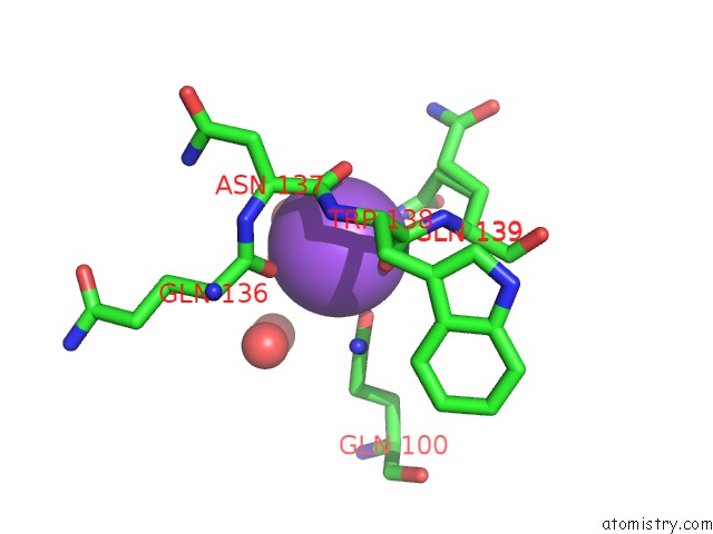
Mono view
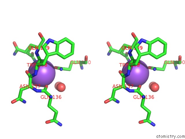
Stereo pair view
Mono view
Stereo pair view
A full contact list of Sodium with other atoms in the Na binding
site number 1 of X-Ray Crystal Structure of Ampc Beta-Lactamase (Ampc(D)) From An Escherichia Coli with A Tripeptide Deletion (GLY286 SER287 ASP288) on the H10 Helix within 5.0Å range:
|
Sodium binding site 2 out of 2 in 2zj9
Go back to
Sodium binding site 2 out
of 2 in the X-Ray Crystal Structure of Ampc Beta-Lactamase (Ampc(D)) From An Escherichia Coli with A Tripeptide Deletion (GLY286 SER287 ASP288) on the H10 Helix
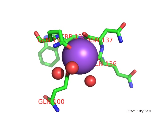
Mono view
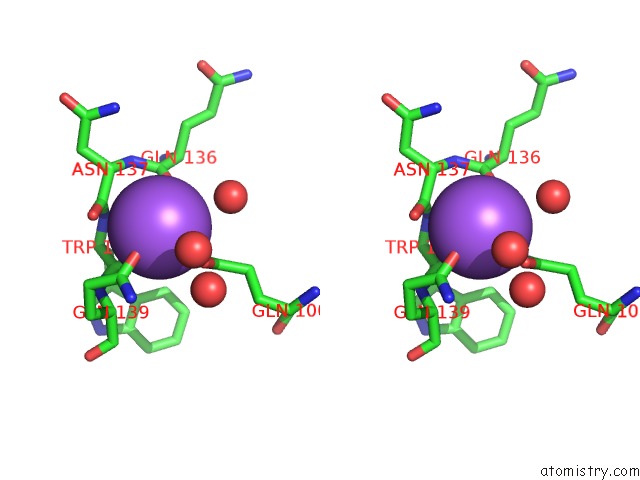
Stereo pair view
Mono view
Stereo pair view
A full contact list of Sodium with other atoms in the Na binding
site number 2 of X-Ray Crystal Structure of Ampc Beta-Lactamase (Ampc(D)) From An Escherichia Coli with A Tripeptide Deletion (GLY286 SER287 ASP288) on the H10 Helix within 5.0Å range:
|
Reference:
Y.Yamaguchi,
G.Sato,
Y.Yamagata,
Y.Doi,
J.Wachino,
Y.Arakawa,
K.Matsuda,
H.Kurosaki.
Structure of Ampc Beta-Lactamase (Ampcd) From An Escherichia Coli Clinical Isolate with A Tripeptide Deletion (GLY286-SER287-ASP288) in the H10 Helix Acta Crystallogr.,Sect.F V. 65 540 2009.
ISSN: ESSN 1744-3091
PubMed: 19478427
DOI: 10.1107/S1744309109014249
Page generated: Sun Aug 17 12:33:27 2025
ISSN: ESSN 1744-3091
PubMed: 19478427
DOI: 10.1107/S1744309109014249
Last articles
Mn in 9LJUMn in 9LJW
Mn in 9LJS
Mn in 9LJR
Mn in 9LJT
Mn in 9LJV
Mg in 9UA2
Mg in 9R96
Mg in 9VM1
Mg in 9P01