Sodium »
PDB 2yfa-2zgb »
2yme »
Sodium in PDB 2yme: Crystal Structure of A Mutant Binding Protein (5HTBP-Achbp) in Complex with Granisetron
Protein crystallography data
The structure of Crystal Structure of A Mutant Binding Protein (5HTBP-Achbp) in Complex with Granisetron, PDB code: 2yme
was solved by
D.Kesters,
A.J.Thompson,
M.Brams,
R.V.Elk,
R.Spurny,
M.Geitmann,
J.M.Villalgordo,
A.Guskov,
U.H.Danielson,
S.C.R.Lummis,
A.B.Smit,
C.Ulens,
with X-Ray Crystallography technique. A brief refinement statistics is given in the table below:
| Resolution Low / High (Å) | 48.908 / 2.40 |
| Space group | P 1 21 1 |
| Cell size a, b, c (Å), α, β, γ (°) | 83.090, 138.900, 119.890, 90.00, 91.07, 90.00 |
| R / Rfree (%) | 16.95 / 22.63 |
Sodium Binding Sites:
The binding sites of Sodium atom in the Crystal Structure of A Mutant Binding Protein (5HTBP-Achbp) in Complex with Granisetron
(pdb code 2yme). This binding sites where shown within
5.0 Angstroms radius around Sodium atom.
In total only one binding site of Sodium was determined in the Crystal Structure of A Mutant Binding Protein (5HTBP-Achbp) in Complex with Granisetron, PDB code: 2yme:
In total only one binding site of Sodium was determined in the Crystal Structure of A Mutant Binding Protein (5HTBP-Achbp) in Complex with Granisetron, PDB code: 2yme:
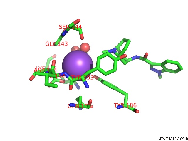
Sodium binding site 1 out of 1 in 2yme
Go back to
Sodium binding site 1 out
of 1 in the Crystal Structure of A Mutant Binding Protein (5HTBP-Achbp) in Complex with Granisetron
Mono view

Stereo pair view
Mono view
Stereo pair view
A full contact list of Sodium with other atoms in the Na binding
site number 1 of Crystal Structure of A Mutant Binding Protein (5HTBP-Achbp) in Complex with Granisetron within 5.0Å range:
|
Reference:
D.Kesters,
A.J.Thompson,
M.Brams,
R.Van Elk,
R.Spurny,
M.Geitmann,
J.M.Villalgordo,
A.Guskov,
U.Helena Danielson,
S.C.R.Lummis,
A.B.Smit,
C.Ulens.
Structural Basis of Ligand Recognition in 5-HT3 Receptors. Embo Rep. V. 14 49 2013.
ISSN: ISSN 1469-221X
PubMed: 23196367
DOI: 10.1038/EMBOR.2012.189
Page generated: Sun Aug 17 12:26:45 2025
ISSN: ISSN 1469-221X
PubMed: 23196367
DOI: 10.1038/EMBOR.2012.189
Last articles
Mn in 9LJUMn in 9LJW
Mn in 9LJS
Mn in 9LJR
Mn in 9LJT
Mn in 9LJV
Mg in 9UA2
Mg in 9R96
Mg in 9VM1
Mg in 9P01