Sodium »
PDB 2x5x-2xjq »
2xhb »
Sodium in PDB 2xhb: Crystal Structure of Dna Polymerase From Thermococcus Gorgonarius in Complex with Hypoxanthine-Containing Dna
Enzymatic activity of Crystal Structure of Dna Polymerase From Thermococcus Gorgonarius in Complex with Hypoxanthine-Containing Dna
All present enzymatic activity of Crystal Structure of Dna Polymerase From Thermococcus Gorgonarius in Complex with Hypoxanthine-Containing Dna:
2.7.7.7;
2.7.7.7;
Protein crystallography data
The structure of Crystal Structure of Dna Polymerase From Thermococcus Gorgonarius in Complex with Hypoxanthine-Containing Dna, PDB code: 2xhb
was solved by
T.Killelea,
S.Ghosh,
S.S.Tan,
P.Heslop,
S.J.Firbank,
E.T.Kool,
B.A.Connolly,
with X-Ray Crystallography technique. A brief refinement statistics is given in the table below:
| Resolution Low / High (Å) | 47.01 / 2.72 |
| Space group | P 21 21 21 |
| Cell size a, b, c (Å), α, β, γ (°) | 79.450, 98.370, 116.630, 90.00, 90.00, 90.00 |
| R / Rfree (%) | 23.361 / 29.455 |
Sodium Binding Sites:
The binding sites of Sodium atom in the Crystal Structure of Dna Polymerase From Thermococcus Gorgonarius in Complex with Hypoxanthine-Containing Dna
(pdb code 2xhb). This binding sites where shown within
5.0 Angstroms radius around Sodium atom.
In total only one binding site of Sodium was determined in the Crystal Structure of Dna Polymerase From Thermococcus Gorgonarius in Complex with Hypoxanthine-Containing Dna, PDB code: 2xhb:
In total only one binding site of Sodium was determined in the Crystal Structure of Dna Polymerase From Thermococcus Gorgonarius in Complex with Hypoxanthine-Containing Dna, PDB code: 2xhb:
Sodium binding site 1 out of 1 in 2xhb
Go back to
Sodium binding site 1 out
of 1 in the Crystal Structure of Dna Polymerase From Thermococcus Gorgonarius in Complex with Hypoxanthine-Containing Dna
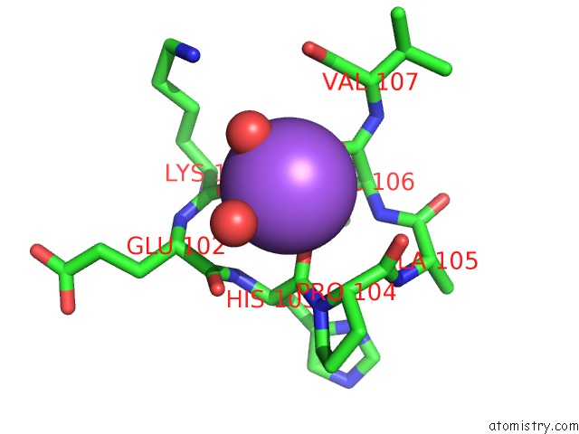
Mono view
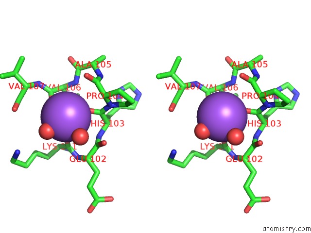
Stereo pair view
Mono view
Stereo pair view
A full contact list of Sodium with other atoms in the Na binding
site number 1 of Crystal Structure of Dna Polymerase From Thermococcus Gorgonarius in Complex with Hypoxanthine-Containing Dna within 5.0Å range:
|
Reference:
T.Killelea,
S.Ghosh,
S.S.Tan,
P.Heslop,
S.J.Firbank,
E.T.Kool,
B.A.Connolly.
Probing the Interaction of Archaeal Dna Polymerases with Deaminated Bases Using X-Ray Crystallography and Non-Hydrogen Bonding Isosteric Base Analogues. Biochemistry V. 49 5772 2010.
ISSN: ISSN 0006-2960
PubMed: 20527806
DOI: 10.1021/BI100421R
Page generated: Mon Oct 7 05:10:29 2024
ISSN: ISSN 0006-2960
PubMed: 20527806
DOI: 10.1021/BI100421R
Last articles
Mg in 3B9BMg in 3B8E
Mg in 3B7L
Mg in 3B6B
Mg in 3B7W
Mg in 3B6V
Mg in 3B6U
Mg in 3B5I
Mg in 3B4A
Mg in 3B6R