Sodium »
PDB 2woi-2x2v »
2ws3 »
Sodium in PDB 2ws3: Crystal Structure of the E. Coli Succinate:Quinone Oxidoreductase (Sqr) Sdhd TYR83PHE Mutant
Enzymatic activity of Crystal Structure of the E. Coli Succinate:Quinone Oxidoreductase (Sqr) Sdhd TYR83PHE Mutant
All present enzymatic activity of Crystal Structure of the E. Coli Succinate:Quinone Oxidoreductase (Sqr) Sdhd TYR83PHE Mutant:
1.3.5.1; 1.3.99.1;
1.3.5.1; 1.3.99.1;
Protein crystallography data
The structure of Crystal Structure of the E. Coli Succinate:Quinone Oxidoreductase (Sqr) Sdhd TYR83PHE Mutant, PDB code: 2ws3
was solved by
J.Ruprecht,
V.Yankovskaya,
E.Maklashina,
S.Iwata,
G.Cecchini,
with X-Ray Crystallography technique. A brief refinement statistics is given in the table below:
| Resolution Low / High (Å) | 49.03 / 3.20 |
| Space group | P 21 21 21 |
| Cell size a, b, c (Å), α, β, γ (°) | 119.850, 184.710, 203.310, 90.00, 90.00, 90.00 |
| R / Rfree (%) | 21.87 / 25.271 |
Other elements in 2ws3:
The structure of Crystal Structure of the E. Coli Succinate:Quinone Oxidoreductase (Sqr) Sdhd TYR83PHE Mutant also contains other interesting chemical elements:
| Iron | (Fe) | 30 atoms |
Sodium Binding Sites:
The binding sites of Sodium atom in the Crystal Structure of the E. Coli Succinate:Quinone Oxidoreductase (Sqr) Sdhd TYR83PHE Mutant
(pdb code 2ws3). This binding sites where shown within
5.0 Angstroms radius around Sodium atom.
In total 3 binding sites of Sodium where determined in the Crystal Structure of the E. Coli Succinate:Quinone Oxidoreductase (Sqr) Sdhd TYR83PHE Mutant, PDB code: 2ws3:
Jump to Sodium binding site number: 1; 2; 3;
In total 3 binding sites of Sodium where determined in the Crystal Structure of the E. Coli Succinate:Quinone Oxidoreductase (Sqr) Sdhd TYR83PHE Mutant, PDB code: 2ws3:
Jump to Sodium binding site number: 1; 2; 3;
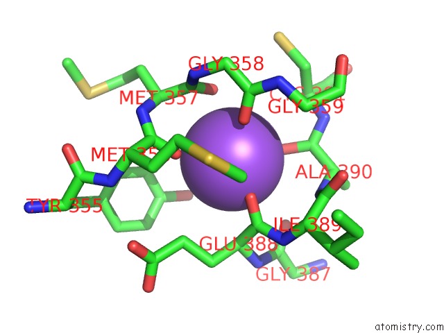
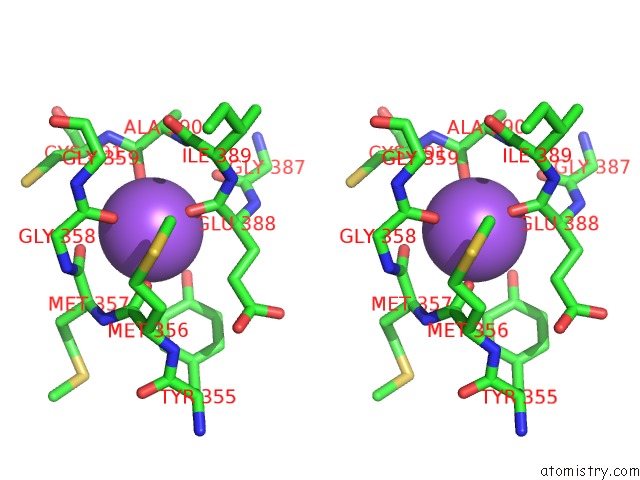
Sodium binding site 1 out of 3 in 2ws3
Go back to
Sodium binding site 1 out
of 3 in the Crystal Structure of the E. Coli Succinate:Quinone Oxidoreductase (Sqr) Sdhd TYR83PHE Mutant
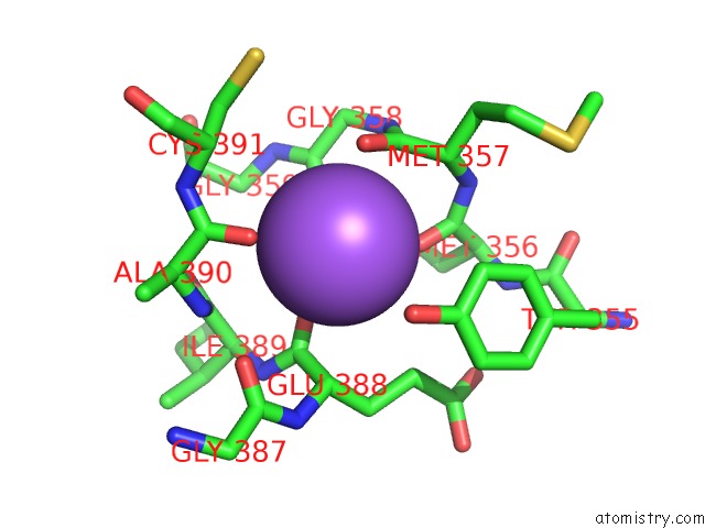
Mono view
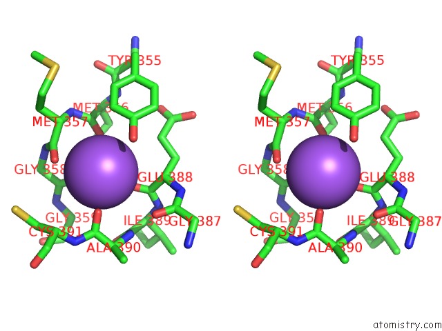
Stereo pair view
Mono view
Stereo pair view
A full contact list of Sodium with other atoms in the Na binding
site number 1 of Crystal Structure of the E. Coli Succinate:Quinone Oxidoreductase (Sqr) Sdhd TYR83PHE Mutant within 5.0Å range:
|
Sodium binding site 2 out of 3 in 2ws3
Go back to
Sodium binding site 2 out
of 3 in the Crystal Structure of the E. Coli Succinate:Quinone Oxidoreductase (Sqr) Sdhd TYR83PHE Mutant
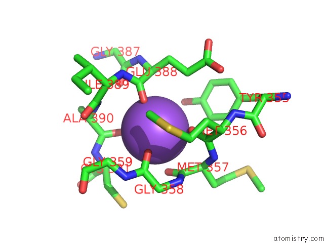
Mono view
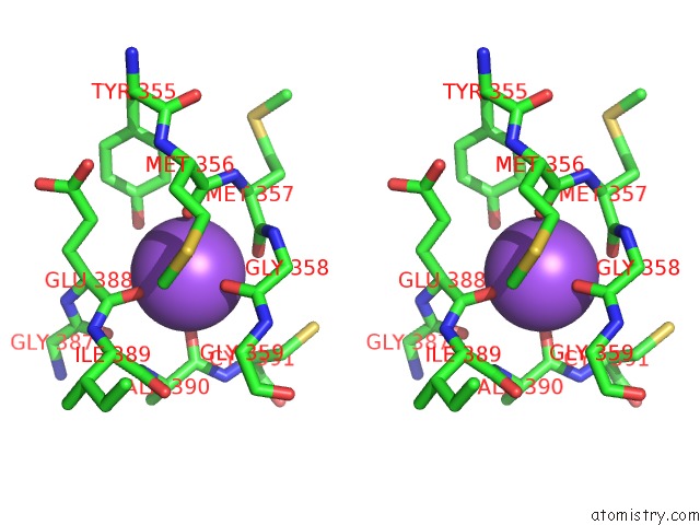
Stereo pair view
Mono view
Stereo pair view
A full contact list of Sodium with other atoms in the Na binding
site number 2 of Crystal Structure of the E. Coli Succinate:Quinone Oxidoreductase (Sqr) Sdhd TYR83PHE Mutant within 5.0Å range:
|
Sodium binding site 3 out of 3 in 2ws3
Go back to
Sodium binding site 3 out
of 3 in the Crystal Structure of the E. Coli Succinate:Quinone Oxidoreductase (Sqr) Sdhd TYR83PHE Mutant
Mono view
Stereo pair view
Mono view
Stereo pair view
A full contact list of Sodium with other atoms in the Na binding
site number 3 of Crystal Structure of the E. Coli Succinate:Quinone Oxidoreductase (Sqr) Sdhd TYR83PHE Mutant within 5.0Å range:
|
Reference:
J.Ruprecht,
V.Yankovskaya,
E.Maklashina,
S.Iwata,
G.Cecchini.
Succinate Dehydrogenase Activity To Be Published.
Page generated: Mon Oct 7 04:55:12 2024
Last articles
Mg in 4X5CMg in 4X5E
Mg in 4X5V
Mg in 4X5B
Mg in 4X59
Mg in 4X58
Mg in 4X4V
Mg in 4X4S
Mg in 4X4R
Mg in 4X4Q