Sodium »
PDB 2wd0-2wof »
2wnn »
Sodium in PDB 2wnn: Structure of Wild Type E. Coli N-Acetylneuraminic Acid Lyase in Complex with Pyruvate in Space Group P21
Enzymatic activity of Structure of Wild Type E. Coli N-Acetylneuraminic Acid Lyase in Complex with Pyruvate in Space Group P21
All present enzymatic activity of Structure of Wild Type E. Coli N-Acetylneuraminic Acid Lyase in Complex with Pyruvate in Space Group P21:
4.1.3.3;
4.1.3.3;
Protein crystallography data
The structure of Structure of Wild Type E. Coli N-Acetylneuraminic Acid Lyase in Complex with Pyruvate in Space Group P21, PDB code: 2wnn
was solved by
I.Campeotto,
A.H.Bolt,
T.A.Harman,
C.H.Trinh,
C.A.Dennis,
S.E.V.Phillips,
A.R.Pearson,
A.Nelson,
A.Berry,
with X-Ray Crystallography technique. A brief refinement statistics is given in the table below:
| Resolution Low / High (Å) | 79.07 / 1.65 |
| Space group | P 1 21 1 |
| Cell size a, b, c (Å), α, β, γ (°) | 54.693, 142.456, 83.626, 90.00, 109.16, 90.00 |
| R / Rfree (%) | 20.986 / 24.92 |
Sodium Binding Sites:
The binding sites of Sodium atom in the Structure of Wild Type E. Coli N-Acetylneuraminic Acid Lyase in Complex with Pyruvate in Space Group P21
(pdb code 2wnn). This binding sites where shown within
5.0 Angstroms radius around Sodium atom.
In total only one binding site of Sodium was determined in the Structure of Wild Type E. Coli N-Acetylneuraminic Acid Lyase in Complex with Pyruvate in Space Group P21, PDB code: 2wnn:
In total only one binding site of Sodium was determined in the Structure of Wild Type E. Coli N-Acetylneuraminic Acid Lyase in Complex with Pyruvate in Space Group P21, PDB code: 2wnn:
Sodium binding site 1 out of 1 in 2wnn
Go back to
Sodium binding site 1 out
of 1 in the Structure of Wild Type E. Coli N-Acetylneuraminic Acid Lyase in Complex with Pyruvate in Space Group P21
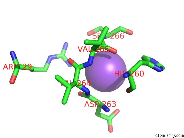
Mono view
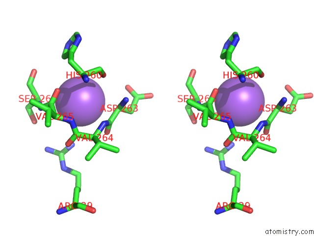
Stereo pair view
Mono view
Stereo pair view
A full contact list of Sodium with other atoms in the Na binding
site number 1 of Structure of Wild Type E. Coli N-Acetylneuraminic Acid Lyase in Complex with Pyruvate in Space Group P21 within 5.0Å range:
|
Reference:
I.Campeotto,
A.H.Bolt,
T.A.Harman,
C.A.Dennis,
C.H.Trinh,
S.E.V.Phillips,
A.Nelson,
A.R.Pearson,
A.Berry.
Structural Insights Into Substrate Specificity in Variants of N-Acetylneuraminic Acid Lyase Produced By Directed Evolution. J.Mol.Biol. V. 404 56 2010.
ISSN: ISSN 0022-2836
PubMed: 20826162
DOI: 10.1016/J.JMB.2010.08.008
Page generated: Mon Oct 7 04:43:20 2024
ISSN: ISSN 0022-2836
PubMed: 20826162
DOI: 10.1016/J.JMB.2010.08.008
Last articles
Zn in 9MJ5Zn in 9HNW
Zn in 9G0L
Zn in 9FNE
Zn in 9DZN
Zn in 9E0I
Zn in 9D32
Zn in 9DAK
Zn in 8ZXC
Zn in 8ZUF