Sodium »
PDB 2qd8-2r22 »
2qw1 »
Sodium in PDB 2qw1: Glucose/Galactose Binding Protein Bound to 3-O-Methyl D-Glucose
Protein crystallography data
The structure of Glucose/Galactose Binding Protein Bound to 3-O-Methyl D-Glucose, PDB code: 2qw1
was solved by
M.J.Borrok,
L.L.Kiessling,
K.T.Forest,
with X-Ray Crystallography technique. A brief refinement statistics is given in the table below:
| Resolution Low / High (Å) | 40.00 / 1.70 |
| Space group | P 21 21 21 |
| Cell size a, b, c (Å), α, β, γ (°) | 56.930, 74.660, 110.070, 90.00, 90.00, 90.00 |
| R / Rfree (%) | 18 / 20.4 |
Other elements in 2qw1:
The structure of Glucose/Galactose Binding Protein Bound to 3-O-Methyl D-Glucose also contains other interesting chemical elements:
| Calcium | (Ca) | 1 atom |
Sodium Binding Sites:
The binding sites of Sodium atom in the Glucose/Galactose Binding Protein Bound to 3-O-Methyl D-Glucose
(pdb code 2qw1). This binding sites where shown within
5.0 Angstroms radius around Sodium atom.
In total 2 binding sites of Sodium where determined in the Glucose/Galactose Binding Protein Bound to 3-O-Methyl D-Glucose, PDB code: 2qw1:
Jump to Sodium binding site number: 1; 2;
In total 2 binding sites of Sodium where determined in the Glucose/Galactose Binding Protein Bound to 3-O-Methyl D-Glucose, PDB code: 2qw1:
Jump to Sodium binding site number: 1; 2;
Sodium binding site 1 out of 2 in 2qw1
Go back to
Sodium binding site 1 out
of 2 in the Glucose/Galactose Binding Protein Bound to 3-O-Methyl D-Glucose
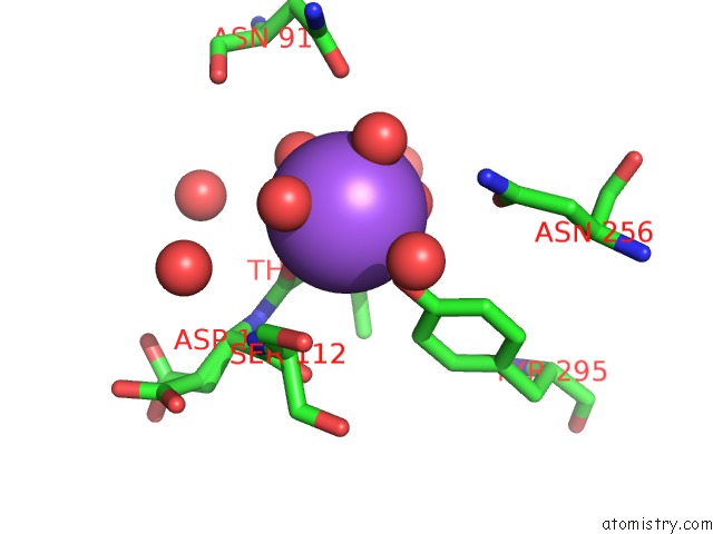
Mono view
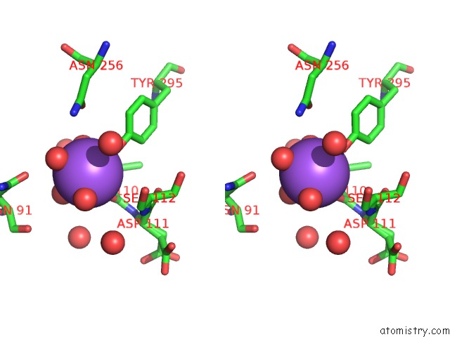
Stereo pair view
Mono view
Stereo pair view
A full contact list of Sodium with other atoms in the Na binding
site number 1 of Glucose/Galactose Binding Protein Bound to 3-O-Methyl D-Glucose within 5.0Å range:
|
Sodium binding site 2 out of 2 in 2qw1
Go back to
Sodium binding site 2 out
of 2 in the Glucose/Galactose Binding Protein Bound to 3-O-Methyl D-Glucose
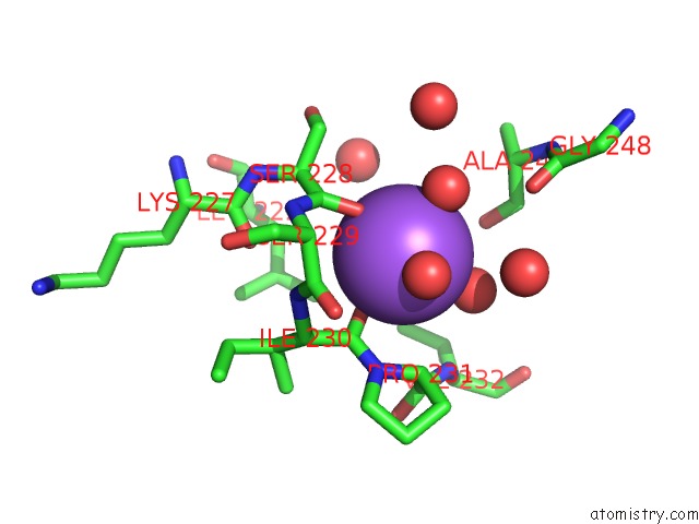
Mono view
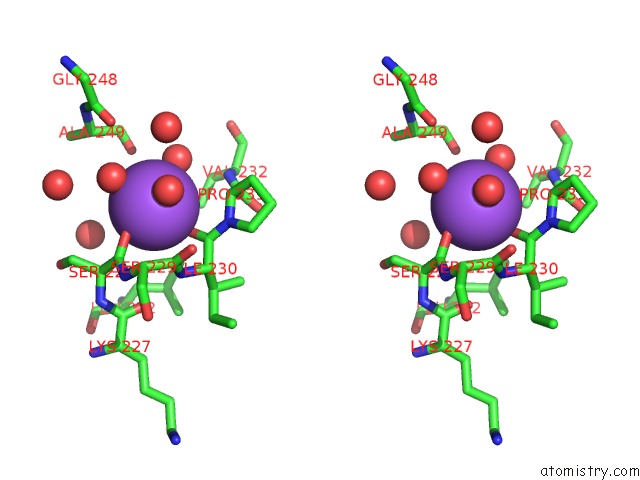
Stereo pair view
Mono view
Stereo pair view
A full contact list of Sodium with other atoms in the Na binding
site number 2 of Glucose/Galactose Binding Protein Bound to 3-O-Methyl D-Glucose within 5.0Å range:
|
Reference:
M.J.Borrok,
Y.Zhu,
K.T.Forest,
L.L.Kiessling.
Structure-Based Design of A Periplasmic Binding Protein Antagonist That Prevents Domain Closure. Acs Chem.Biol. V. 4 447 2009.
ISSN: ISSN 1554-8929
PubMed: 19348466
DOI: 10.1021/CB900021Q
Page generated: Mon Oct 7 04:04:29 2024
ISSN: ISSN 1554-8929
PubMed: 19348466
DOI: 10.1021/CB900021Q
Last articles
Mg in 5IVGMg in 5IUL
Mg in 5IUM
Mg in 5IRP
Mg in 5IUK
Mg in 5IUJ
Mg in 5IUC
Mg in 5IU0
Mg in 5IT5
Mg in 5ITZ