Sodium »
PDB 2p0o-2plo »
2pfl »
Sodium in PDB 2pfl: Crystal Structure of Pfl From E.Coli
Enzymatic activity of Crystal Structure of Pfl From E.Coli
All present enzymatic activity of Crystal Structure of Pfl From E.Coli:
2.3.1.54;
2.3.1.54;
Protein crystallography data
The structure of Crystal Structure of Pfl From E.Coli, PDB code: 2pfl
was solved by
A.Becker,
K.Fritz-Wolf,
W.Kabsch,
J.Knappe,
S.Schultz,
A.F.V.Wagner,
with X-Ray Crystallography technique. A brief refinement statistics is given in the table below:
| Resolution Low / High (Å) | 15.00 / 2.90 |
| Space group | P 43 21 2 |
| Cell size a, b, c (Å), α, β, γ (°) | 159.020, 159.020, 160.640, 90.00, 90.00, 90.00 |
| R / Rfree (%) | 20.2 / 24.2 |
Other elements in 2pfl:
The structure of Crystal Structure of Pfl From E.Coli also contains other interesting chemical elements:
| Chlorine | (Cl) | 2 atoms |
Sodium Binding Sites:
The binding sites of Sodium atom in the Crystal Structure of Pfl From E.Coli
(pdb code 2pfl). This binding sites where shown within
5.0 Angstroms radius around Sodium atom.
In total 2 binding sites of Sodium where determined in the Crystal Structure of Pfl From E.Coli, PDB code: 2pfl:
Jump to Sodium binding site number: 1; 2;
In total 2 binding sites of Sodium where determined in the Crystal Structure of Pfl From E.Coli, PDB code: 2pfl:
Jump to Sodium binding site number: 1; 2;
Sodium binding site 1 out of 2 in 2pfl
Go back to
Sodium binding site 1 out
of 2 in the Crystal Structure of Pfl From E.Coli

Mono view
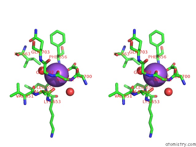
Stereo pair view
Mono view
Stereo pair view
A full contact list of Sodium with other atoms in the Na binding
site number 1 of Crystal Structure of Pfl From E.Coli within 5.0Å range:
|
Sodium binding site 2 out of 2 in 2pfl
Go back to
Sodium binding site 2 out
of 2 in the Crystal Structure of Pfl From E.Coli
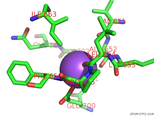
Mono view
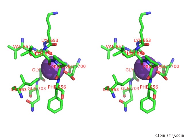
Stereo pair view
Mono view
Stereo pair view
A full contact list of Sodium with other atoms in the Na binding
site number 2 of Crystal Structure of Pfl From E.Coli within 5.0Å range:
|
Reference:
A.Becker,
K.Fritz-Wolf,
W.Kabsch,
J.Knappe,
S.Schultz,
A.F.Volker Wagner.
Structure and Mechanism of the Glycyl Radical Enzyme Pyruvate Formate-Lyase. Nat.Struct.Biol. V. 6 969 1999.
ISSN: ISSN 1072-8368
PubMed: 10504733
DOI: 10.1038/13341
Page generated: Mon Oct 7 03:41:13 2024
ISSN: ISSN 1072-8368
PubMed: 10504733
DOI: 10.1038/13341
Last articles
I in 4AGMI in 4AGL
I in 4AGN
I in 464D
I in 4A3P
I in 444D
I in 445D
I in 449D
I in 448D
I in 442D