Sodium »
PDB 2ok7-2ozt »
2oyc »
Sodium in PDB 2oyc: Crystal Structure of Human Pyridoxal Phosphate Phosphatase
Enzymatic activity of Crystal Structure of Human Pyridoxal Phosphate Phosphatase
All present enzymatic activity of Crystal Structure of Human Pyridoxal Phosphate Phosphatase:
3.1.3.74;
3.1.3.74;
Protein crystallography data
The structure of Crystal Structure of Human Pyridoxal Phosphate Phosphatase, PDB code: 2oyc
was solved by
U.A.Ramagopal,
J.Freeman,
M.Izuka,
R.Toro,
J.M.Sauder,
S.K.Burley,
S.C.Almo,
New York Sgx Research Center For Structural Genomics(Nysgxrc),
with X-Ray Crystallography technique. A brief refinement statistics is given in the table below:
| Resolution Low / High (Å) | 26.90 / 1.72 |
| Space group | P 43 21 2 |
| Cell size a, b, c (Å), α, β, γ (°) | 54.329, 54.329, 213.200, 90.00, 90.00, 90.00 |
| R / Rfree (%) | 19.2 / 22.3 |
Other elements in 2oyc:
The structure of Crystal Structure of Human Pyridoxal Phosphate Phosphatase also contains other interesting chemical elements:
| Tungsten | (W) | 1 atom |
Sodium Binding Sites:
The binding sites of Sodium atom in the Crystal Structure of Human Pyridoxal Phosphate Phosphatase
(pdb code 2oyc). This binding sites where shown within
5.0 Angstroms radius around Sodium atom.
In total only one binding site of Sodium was determined in the Crystal Structure of Human Pyridoxal Phosphate Phosphatase, PDB code: 2oyc:
In total only one binding site of Sodium was determined in the Crystal Structure of Human Pyridoxal Phosphate Phosphatase, PDB code: 2oyc:
Sodium binding site 1 out of 1 in 2oyc
Go back to
Sodium binding site 1 out
of 1 in the Crystal Structure of Human Pyridoxal Phosphate Phosphatase
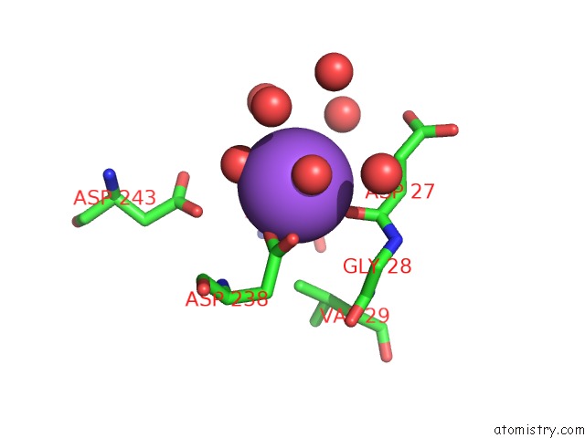
Mono view
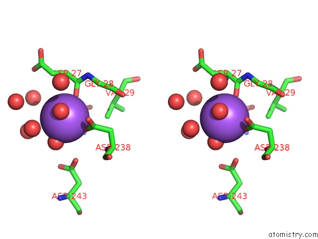
Stereo pair view
Mono view
Stereo pair view
A full contact list of Sodium with other atoms in the Na binding
site number 1 of Crystal Structure of Human Pyridoxal Phosphate Phosphatase within 5.0Å range:
|
Reference:
S.C.Almo,
J.B.Bonanno,
J.M.Sauder,
S.Emtage,
T.P.Dilorenzo,
V.Malashkevich,
S.R.Wasserman,
S.Swaminathan,
S.Eswaramoorthy,
R.Agarwal,
D.Kumaran,
M.Madegowda,
S.Ragumani,
Y.Patskovsky,
J.Alvarado,
U.A.Ramagopal,
J.Faber-Barata,
M.R.Chance,
A.Sali,
A.Fiser,
Z.Y.Zhang,
D.S.Lawrence,
S.K.Burley.
Structural Genomics of Protein Phosphatases. J.Struct.Funct.Genom. V. 8 121 2007.
ISSN: ISSN 1345-711X
PubMed: 18058037
DOI: 10.1007/S10969-007-9036-1
Page generated: Sun Aug 17 11:06:25 2025
ISSN: ISSN 1345-711X
PubMed: 18058037
DOI: 10.1007/S10969-007-9036-1
Last articles
K in 9NESK in 9PHG
K in 9NEI
K in 9NED
K in 9NEC
K in 9NEG
K in 9CWU
K in 9CVB
K in 9CVA
K in 9COM