Sodium »
PDB 2fmp-2gg2 »
2g79 »
Sodium in PDB 2g79: Crystal Structure of the R132K:Y134F Mutant of Cellular Retinoic Acid Binding Protein Type II in Complex with All-Trans-Retinal at 1.69 Angstroms Resolution
Protein crystallography data
The structure of Crystal Structure of the R132K:Y134F Mutant of Cellular Retinoic Acid Binding Protein Type II in Complex with All-Trans-Retinal at 1.69 Angstroms Resolution, PDB code: 2g79
was solved by
S.Vaezeslami,
J.H.Geiger,
with X-Ray Crystallography technique. A brief refinement statistics is given in the table below:
| Resolution Low / High (Å) | 39.19 / 1.69 |
| Space group | P 21 21 21 |
| Cell size a, b, c (Å), α, β, γ (°) | 45.493, 46.325, 73.597, 90.00, 90.00, 90.00 |
| R / Rfree (%) | 15.5 / 21.4 |
Sodium Binding Sites:
The binding sites of Sodium atom in the Crystal Structure of the R132K:Y134F Mutant of Cellular Retinoic Acid Binding Protein Type II in Complex with All-Trans-Retinal at 1.69 Angstroms Resolution
(pdb code 2g79). This binding sites where shown within
5.0 Angstroms radius around Sodium atom.
In total 4 binding sites of Sodium where determined in the Crystal Structure of the R132K:Y134F Mutant of Cellular Retinoic Acid Binding Protein Type II in Complex with All-Trans-Retinal at 1.69 Angstroms Resolution, PDB code: 2g79:
Jump to Sodium binding site number: 1; 2; 3; 4;
In total 4 binding sites of Sodium where determined in the Crystal Structure of the R132K:Y134F Mutant of Cellular Retinoic Acid Binding Protein Type II in Complex with All-Trans-Retinal at 1.69 Angstroms Resolution, PDB code: 2g79:
Jump to Sodium binding site number: 1; 2; 3; 4;
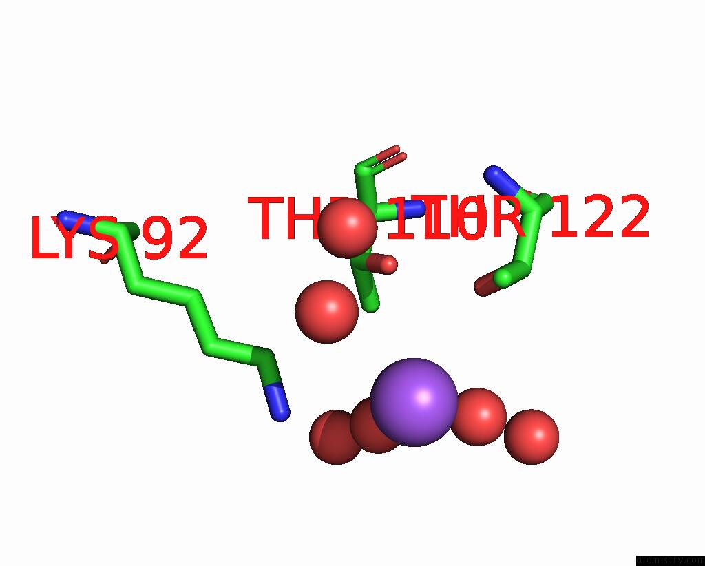
Sodium binding site 1 out of 4 in 2g79
Go back to
Sodium binding site 1 out
of 4 in the Crystal Structure of the R132K:Y134F Mutant of Cellular Retinoic Acid Binding Protein Type II in Complex with All-Trans-Retinal at 1.69 Angstroms Resolution
Mono view

Stereo pair view
Mono view
Stereo pair view
A full contact list of Sodium with other atoms in the Na binding
site number 1 of Crystal Structure of the R132K:Y134F Mutant of Cellular Retinoic Acid Binding Protein Type II in Complex with All-Trans-Retinal at 1.69 Angstroms Resolution within 5.0Å range:
|
Sodium binding site 2 out of 4 in 2g79
Go back to
Sodium binding site 2 out
of 4 in the Crystal Structure of the R132K:Y134F Mutant of Cellular Retinoic Acid Binding Protein Type II in Complex with All-Trans-Retinal at 1.69 Angstroms Resolution

Mono view

Stereo pair view
Mono view
Stereo pair view
A full contact list of Sodium with other atoms in the Na binding
site number 2 of Crystal Structure of the R132K:Y134F Mutant of Cellular Retinoic Acid Binding Protein Type II in Complex with All-Trans-Retinal at 1.69 Angstroms Resolution within 5.0Å range:
|
Sodium binding site 3 out of 4 in 2g79
Go back to
Sodium binding site 3 out
of 4 in the Crystal Structure of the R132K:Y134F Mutant of Cellular Retinoic Acid Binding Protein Type II in Complex with All-Trans-Retinal at 1.69 Angstroms Resolution

Mono view

Stereo pair view
Mono view
Stereo pair view
A full contact list of Sodium with other atoms in the Na binding
site number 3 of Crystal Structure of the R132K:Y134F Mutant of Cellular Retinoic Acid Binding Protein Type II in Complex with All-Trans-Retinal at 1.69 Angstroms Resolution within 5.0Å range:
|
Sodium binding site 4 out of 4 in 2g79
Go back to
Sodium binding site 4 out
of 4 in the Crystal Structure of the R132K:Y134F Mutant of Cellular Retinoic Acid Binding Protein Type II in Complex with All-Trans-Retinal at 1.69 Angstroms Resolution

Mono view

Stereo pair view
Mono view
Stereo pair view
A full contact list of Sodium with other atoms in the Na binding
site number 4 of Crystal Structure of the R132K:Y134F Mutant of Cellular Retinoic Acid Binding Protein Type II in Complex with All-Trans-Retinal at 1.69 Angstroms Resolution within 5.0Å range:
|
Reference:
C.Vasileiou,
S.Vaezeslami,
R.M.Crist,
M.Rabago-Smith,
J.H.Geiger,
B.Borhan.
Protein Design: Reengineering Cellular Retinoic Acid Binding Protein II Into A Rhodopsin Protein Mimic. J.Am.Chem.Soc. V. 129 6140 2007.
ISSN: ISSN 0002-7863
PubMed: 17447762
DOI: 10.1021/JA067546R
Page generated: Mon Oct 7 02:32:36 2024
ISSN: ISSN 0002-7863
PubMed: 17447762
DOI: 10.1021/JA067546R
Last articles
I in 5M0MI in 5M7M
I in 5M0E
I in 5M0D
I in 5LQQ
I in 5L1A
I in 5LBD
I in 5LZK
I in 5KST
I in 5L3J