Sodium »
PDB 2czs-2e4r »
2dv3 »
Sodium in PDB 2dv3: Crystal Structure of LEU65 to Arg Mutant of Diphthine Synthase
Enzymatic activity of Crystal Structure of LEU65 to Arg Mutant of Diphthine Synthase
All present enzymatic activity of Crystal Structure of LEU65 to Arg Mutant of Diphthine Synthase:
2.1.1.98;
2.1.1.98;
Protein crystallography data
The structure of Crystal Structure of LEU65 to Arg Mutant of Diphthine Synthase, PDB code: 2dv3
was solved by
H.Mizutani,
Y.Matsuura,
N.Kunishima,
Riken Structuralgenomics/Proteomics Initiative (Rsgi),
with X-Ray Crystallography technique. A brief refinement statistics is given in the table below:
| Resolution Low / High (Å) | 30.00 / 1.90 |
| Space group | P 41 21 2 |
| Cell size a, b, c (Å), α, β, γ (°) | 104.197, 104.197, 137.827, 90.00, 90.00, 90.00 |
| R / Rfree (%) | 21.9 / 23.4 |
Other elements in 2dv3:
The structure of Crystal Structure of LEU65 to Arg Mutant of Diphthine Synthase also contains other interesting chemical elements:
| Chlorine | (Cl) | 1 atom |
Sodium Binding Sites:
The binding sites of Sodium atom in the Crystal Structure of LEU65 to Arg Mutant of Diphthine Synthase
(pdb code 2dv3). This binding sites where shown within
5.0 Angstroms radius around Sodium atom.
In total only one binding site of Sodium was determined in the Crystal Structure of LEU65 to Arg Mutant of Diphthine Synthase, PDB code: 2dv3:
In total only one binding site of Sodium was determined in the Crystal Structure of LEU65 to Arg Mutant of Diphthine Synthase, PDB code: 2dv3:
Sodium binding site 1 out of 1 in 2dv3
Go back to
Sodium binding site 1 out
of 1 in the Crystal Structure of LEU65 to Arg Mutant of Diphthine Synthase
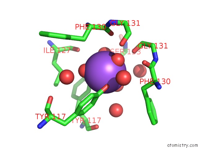
Mono view
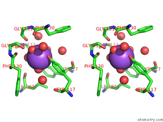
Stereo pair view
Mono view
Stereo pair view
A full contact list of Sodium with other atoms in the Na binding
site number 1 of Crystal Structure of LEU65 to Arg Mutant of Diphthine Synthase within 5.0Å range:
|
Reference:
H.Mizutani,
Y.Matsuura,
N.Kunishima.
Crystal Structure of Diphthine Synthase From Pyrococcus Horikoshii OT3 To Be Published.
Page generated: Mon Oct 7 02:15:51 2024
Last articles
Mg in 4L9ZMg in 4L9Y
Mg in 4LA6
Mg in 4L9W
Mg in 4L81
Mg in 4L9S
Mg in 4L8N
Mg in 4L87
Mg in 4L8G
Mg in 4L80