Sodium »
PDB 2aoe-2bhp »
2asf »
Sodium in PDB 2asf: Crystal Structure of the Conserved Hypothetical Protein RV2074 From Mycobacterium Tuberculosis 1.6 A
Protein crystallography data
The structure of Crystal Structure of the Conserved Hypothetical Protein RV2074 From Mycobacterium Tuberculosis 1.6 A, PDB code: 2asf
was solved by
B.K.Biswal,
K.Au,
M.M.Cherney,
C.Garen,
M.N.James,
Tb Structural Genomicsconsortium (Tbsgc),
with X-Ray Crystallography technique. A brief refinement statistics is given in the table below:
| Resolution Low / High (Å) | 38.24 / 1.60 |
| Space group | P 43 21 2 |
| Cell size a, b, c (Å), α, β, γ (°) | 73.395, 73.395, 44.794, 90.00, 90.00, 90.00 |
| R / Rfree (%) | 17.9 / 20.4 |
Sodium Binding Sites:
The binding sites of Sodium atom in the Crystal Structure of the Conserved Hypothetical Protein RV2074 From Mycobacterium Tuberculosis 1.6 A
(pdb code 2asf). This binding sites where shown within
5.0 Angstroms radius around Sodium atom.
In total only one binding site of Sodium was determined in the Crystal Structure of the Conserved Hypothetical Protein RV2074 From Mycobacterium Tuberculosis 1.6 A, PDB code: 2asf:
In total only one binding site of Sodium was determined in the Crystal Structure of the Conserved Hypothetical Protein RV2074 From Mycobacterium Tuberculosis 1.6 A, PDB code: 2asf:
Sodium binding site 1 out of 1 in 2asf
Go back to
Sodium binding site 1 out
of 1 in the Crystal Structure of the Conserved Hypothetical Protein RV2074 From Mycobacterium Tuberculosis 1.6 A
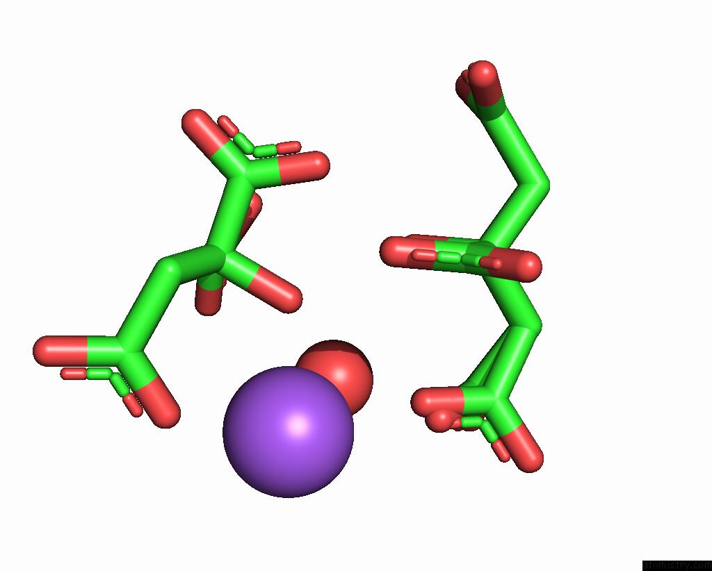
Mono view
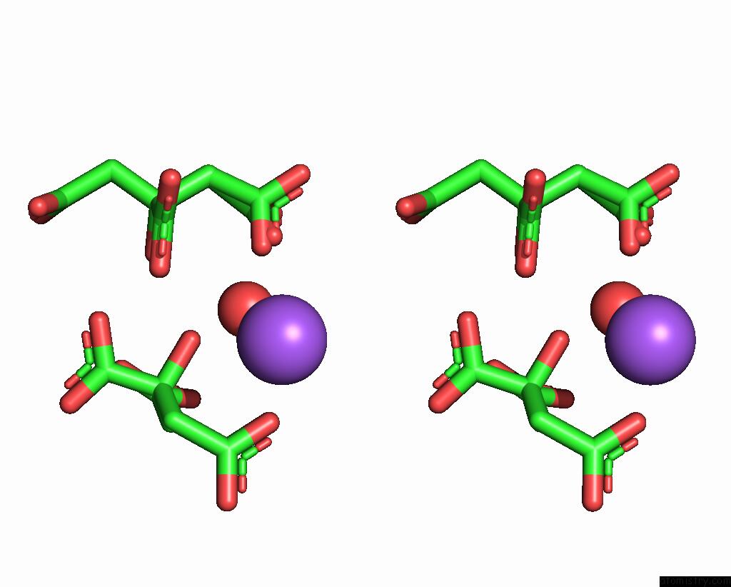
Stereo pair view
Mono view
Stereo pair view
A full contact list of Sodium with other atoms in the Na binding
site number 1 of Crystal Structure of the Conserved Hypothetical Protein RV2074 From Mycobacterium Tuberculosis 1.6 A within 5.0Å range:
|
Reference:
B.K.Biswal,
K.Au,
M.M.Cherney,
C.Garen,
M.N.James.
The Molecular Structure of RV2074, A Probable Pyridoxine 5'-Phosphate Oxidase From Mycobacterium Tuberculosis, at 1.6 Angstroms Resolution. Acta Crystallogr.,Sect.F V. 62 735 2006.
ISSN: ESSN 1744-3091
PubMed: 16880544
DOI: 10.1107/S1744309106025012
Page generated: Mon Oct 7 01:54:37 2024
ISSN: ESSN 1744-3091
PubMed: 16880544
DOI: 10.1107/S1744309106025012
Last articles
Fe in 2YXOFe in 2YRS
Fe in 2YXC
Fe in 2YNM
Fe in 2YVJ
Fe in 2YP1
Fe in 2YU2
Fe in 2YU1
Fe in 2YQB
Fe in 2YOO