Sodium »
PDB 1zum-2aoc »
2a9y »
Sodium in PDB 2a9y: Crystal Structure of T. Gondii Adenosine Kinase Complexed with N6- Dimethyladenosine
Enzymatic activity of Crystal Structure of T. Gondii Adenosine Kinase Complexed with N6- Dimethyladenosine
All present enzymatic activity of Crystal Structure of T. Gondii Adenosine Kinase Complexed with N6- Dimethyladenosine:
2.7.1.20;
2.7.1.20;
Protein crystallography data
The structure of Crystal Structure of T. Gondii Adenosine Kinase Complexed with N6- Dimethyladenosine, PDB code: 2a9y
was solved by
Y.Zhang,
M.H.El Kouni,
S.E.Ealick,
with X-Ray Crystallography technique. A brief refinement statistics is given in the table below:
| Resolution Low / High (Å) | 10.00 / 1.35 |
| Space group | P 21 21 21 |
| Cell size a, b, c (Å), α, β, γ (°) | 60.285, 61.516, 91.828, 90.00, 90.00, 90.00 |
| R / Rfree (%) | 13.4 / 19.6 |
Other elements in 2a9y:
The structure of Crystal Structure of T. Gondii Adenosine Kinase Complexed with N6- Dimethyladenosine also contains other interesting chemical elements:
| Chlorine | (Cl) | 1 atom |
Sodium Binding Sites:
The binding sites of Sodium atom in the Crystal Structure of T. Gondii Adenosine Kinase Complexed with N6- Dimethyladenosine
(pdb code 2a9y). This binding sites where shown within
5.0 Angstroms radius around Sodium atom.
In total 2 binding sites of Sodium where determined in the Crystal Structure of T. Gondii Adenosine Kinase Complexed with N6- Dimethyladenosine, PDB code: 2a9y:
Jump to Sodium binding site number: 1; 2;
In total 2 binding sites of Sodium where determined in the Crystal Structure of T. Gondii Adenosine Kinase Complexed with N6- Dimethyladenosine, PDB code: 2a9y:
Jump to Sodium binding site number: 1; 2;
Sodium binding site 1 out of 2 in 2a9y
Go back to
Sodium binding site 1 out
of 2 in the Crystal Structure of T. Gondii Adenosine Kinase Complexed with N6- Dimethyladenosine
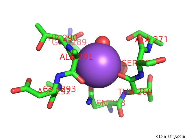
Mono view
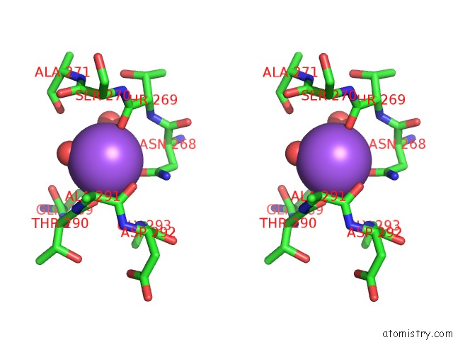
Stereo pair view
Mono view
Stereo pair view
A full contact list of Sodium with other atoms in the Na binding
site number 1 of Crystal Structure of T. Gondii Adenosine Kinase Complexed with N6- Dimethyladenosine within 5.0Å range:
|
Sodium binding site 2 out of 2 in 2a9y
Go back to
Sodium binding site 2 out
of 2 in the Crystal Structure of T. Gondii Adenosine Kinase Complexed with N6- Dimethyladenosine
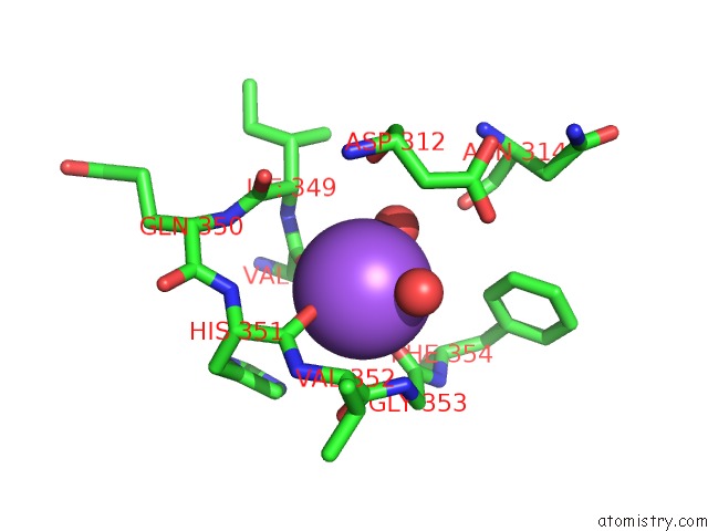
Mono view
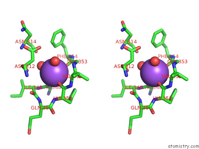
Stereo pair view
Mono view
Stereo pair view
A full contact list of Sodium with other atoms in the Na binding
site number 2 of Crystal Structure of T. Gondii Adenosine Kinase Complexed with N6- Dimethyladenosine within 5.0Å range:
|
Reference:
Y.Zhang,
M.H.El Kouni,
S.E.Ealick.
Substrate Analogs Induce An Intermediate Conformational Change in Toxoplasma Gondii Adenosine Kinase Acta Crystallogr.,Sect.D V. 63 126 2007.
ISSN: ISSN 0907-4449
PubMed: 17242506
DOI: 10.1107/S0907444906043654
Page generated: Mon Oct 7 01:49:58 2024
ISSN: ISSN 0907-4449
PubMed: 17242506
DOI: 10.1107/S0907444906043654
Last articles
K in 1C7JK in 1C35
K in 1BXR
K in 1A9X
K in 1C1D
K in 1C1X
K in 1BW9
K in 1BXG
K in 1BUP
K in 1BL8