Sodium »
PDB 1x9j-1y4d »
1y38 »
Sodium in PDB 1y38: Crystal Structure of the Complex Formed Between Phospholipase A2 Dimer and Glycerophosphate at 2.4 A Resolution
Enzymatic activity of Crystal Structure of the Complex Formed Between Phospholipase A2 Dimer and Glycerophosphate at 2.4 A Resolution
All present enzymatic activity of Crystal Structure of the Complex Formed Between Phospholipase A2 Dimer and Glycerophosphate at 2.4 A Resolution:
3.1.1.4;
3.1.1.4;
Protein crystallography data
The structure of Crystal Structure of the Complex Formed Between Phospholipase A2 Dimer and Glycerophosphate at 2.4 A Resolution, PDB code: 1y38
was solved by
N.Singh,
T.Jabeen,
S.Sharma,
T.P.Singh,
with X-Ray Crystallography technique. A brief refinement statistics is given in the table below:
| Resolution Low / High (Å) | 19.86 / 2.44 |
| Space group | P 21 21 21 |
| Cell size a, b, c (Å), α, β, γ (°) | 45.866, 69.071, 75.646, 90.00, 90.00, 90.00 |
| R / Rfree (%) | 19.4 / 22.4 |
Sodium Binding Sites:
The binding sites of Sodium atom in the Crystal Structure of the Complex Formed Between Phospholipase A2 Dimer and Glycerophosphate at 2.4 A Resolution
(pdb code 1y38). This binding sites where shown within
5.0 Angstroms radius around Sodium atom.
In total only one binding site of Sodium was determined in the Crystal Structure of the Complex Formed Between Phospholipase A2 Dimer and Glycerophosphate at 2.4 A Resolution, PDB code: 1y38:
In total only one binding site of Sodium was determined in the Crystal Structure of the Complex Formed Between Phospholipase A2 Dimer and Glycerophosphate at 2.4 A Resolution, PDB code: 1y38:
Sodium binding site 1 out of 1 in 1y38
Go back to
Sodium binding site 1 out
of 1 in the Crystal Structure of the Complex Formed Between Phospholipase A2 Dimer and Glycerophosphate at 2.4 A Resolution
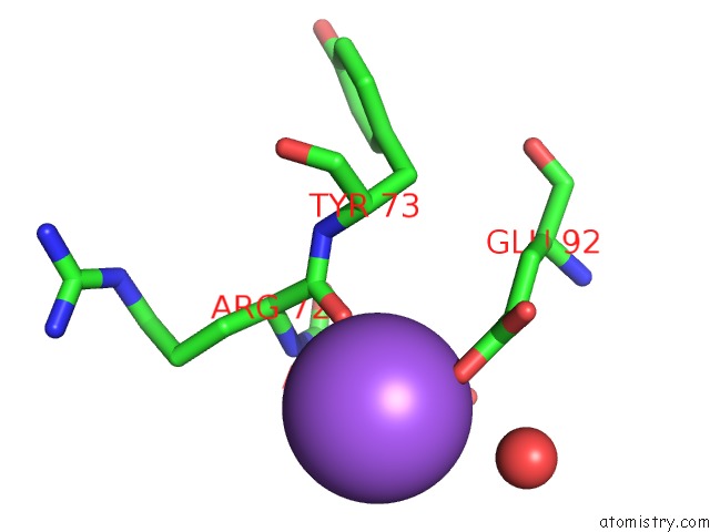
Mono view
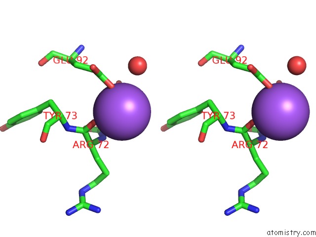
Stereo pair view
Mono view
Stereo pair view
A full contact list of Sodium with other atoms in the Na binding
site number 1 of Crystal Structure of the Complex Formed Between Phospholipase A2 Dimer and Glycerophosphate at 2.4 A Resolution within 5.0Å range:
|
Reference:
N.Singh,
T.Jabeen,
S.Sharma,
T.P.Singh.
Crystal Structure of the Complex Formed Between Phospholipase A2 Dimer and Glycerophosphate at 2.4 A Resolution To Be Published.
Page generated: Mon Oct 7 00:36:33 2024
Last articles
Mg in 5LXIMg in 5LXM
Mg in 5LUF
Mg in 5LWK
Mg in 5LUK
Mg in 5LUI
Mg in 5LU5
Mg in 5LMU
Mg in 5LTT
Mg in 5LU4