Sodium »
PDB 1qtk-1s3x »
1s0a »
Sodium in PDB 1s0a: Crystal Structure of the Y17F Mutant of 7,8-Diaminopelargonic Acid Synthase
Enzymatic activity of Crystal Structure of the Y17F Mutant of 7,8-Diaminopelargonic Acid Synthase
All present enzymatic activity of Crystal Structure of the Y17F Mutant of 7,8-Diaminopelargonic Acid Synthase:
2.6.1.62;
2.6.1.62;
Protein crystallography data
The structure of Crystal Structure of the Y17F Mutant of 7,8-Diaminopelargonic Acid Synthase, PDB code: 1s0a
was solved by
J.Sandmark,
A.C.Eliot,
K.Famm,
G.Schneider,
J.F.Kirsch,
with X-Ray Crystallography technique. A brief refinement statistics is given in the table below:
| Resolution Low / High (Å) | 19.92 / 1.71 |
| Space group | P 1 21 1 |
| Cell size a, b, c (Å), α, β, γ (°) | 58.508, 55.659, 121.396, 90.00, 97.04, 90.00 |
| R / Rfree (%) | 18.6 / 20.6 |
Sodium Binding Sites:
The binding sites of Sodium atom in the Crystal Structure of the Y17F Mutant of 7,8-Diaminopelargonic Acid Synthase
(pdb code 1s0a). This binding sites where shown within
5.0 Angstroms radius around Sodium atom.
In total 2 binding sites of Sodium where determined in the Crystal Structure of the Y17F Mutant of 7,8-Diaminopelargonic Acid Synthase, PDB code: 1s0a:
Jump to Sodium binding site number: 1; 2;
In total 2 binding sites of Sodium where determined in the Crystal Structure of the Y17F Mutant of 7,8-Diaminopelargonic Acid Synthase, PDB code: 1s0a:
Jump to Sodium binding site number: 1; 2;
Sodium binding site 1 out of 2 in 1s0a
Go back to
Sodium binding site 1 out
of 2 in the Crystal Structure of the Y17F Mutant of 7,8-Diaminopelargonic Acid Synthase
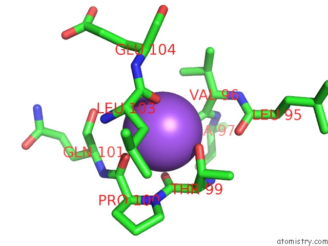
Mono view
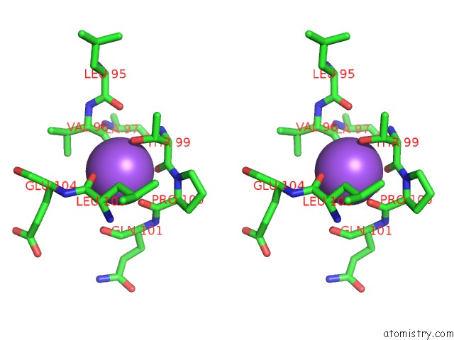
Stereo pair view
Mono view
Stereo pair view
A full contact list of Sodium with other atoms in the Na binding
site number 1 of Crystal Structure of the Y17F Mutant of 7,8-Diaminopelargonic Acid Synthase within 5.0Å range:
|
Sodium binding site 2 out of 2 in 1s0a
Go back to
Sodium binding site 2 out
of 2 in the Crystal Structure of the Y17F Mutant of 7,8-Diaminopelargonic Acid Synthase
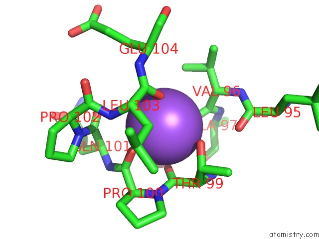
Mono view
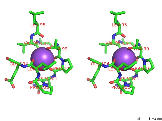
Stereo pair view
Mono view
Stereo pair view
A full contact list of Sodium with other atoms in the Na binding
site number 2 of Crystal Structure of the Y17F Mutant of 7,8-Diaminopelargonic Acid Synthase within 5.0Å range:
|
Reference:
J.Sandmark,
A.C.Eliot,
K.Famm,
G.Schneider,
J.F.Kirsch.
Conserved and Nonconserved Residues in the Substrate Binding Site of 7,8-Diaminopelargonic Acid Synthase From Escherichia Coli Are Essential For Catalysis. Biochemistry V. 43 1213 2004.
ISSN: ISSN 0006-2960
PubMed: 14756557
DOI: 10.1021/BI0358059
Page generated: Sun Aug 17 07:34:49 2025
ISSN: ISSN 0006-2960
PubMed: 14756557
DOI: 10.1021/BI0358059
Last articles
Mn in 9LJUMn in 9LJW
Mn in 9LJS
Mn in 9LJR
Mn in 9LJT
Mn in 9LJV
Mg in 9UA2
Mg in 9R96
Mg in 9VM1
Mg in 9P01