Sodium »
PDB 1ph7-1qsx »
1q93 »
Sodium in PDB 1q93: Crystal Structure of A Mutant of the Sarcin/Ricin Domain From Rat 28S Rrna
Protein crystallography data
The structure of Crystal Structure of A Mutant of the Sarcin/Ricin Domain From Rat 28S Rrna, PDB code: 1q93
was solved by
C.C.Correll,
J.Beneken,
M.J.Plantinga,
M.Lubbers,
Y.L.Chan,
with X-Ray Crystallography technique. A brief refinement statistics is given in the table below:
| Resolution Low / High (Å) | 40.00 / 2.25 |
| Space group | P 43 2 2 |
| Cell size a, b, c (Å), α, β, γ (°) | 42.143, 42.143, 336.167, 90.00, 90.00, 90.00 |
| R / Rfree (%) | 22.5 / 26.6 |
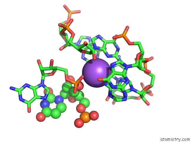
Sodium Binding Sites:
The binding sites of Sodium atom in the Crystal Structure of A Mutant of the Sarcin/Ricin Domain From Rat 28S Rrna
(pdb code 1q93). This binding sites where shown within
5.0 Angstroms radius around Sodium atom.
In total only one binding site of Sodium was determined in the Crystal Structure of A Mutant of the Sarcin/Ricin Domain From Rat 28S Rrna, PDB code: 1q93:
In total only one binding site of Sodium was determined in the Crystal Structure of A Mutant of the Sarcin/Ricin Domain From Rat 28S Rrna, PDB code: 1q93:
Sodium binding site 1 out of 1 in 1q93
Go back to
Sodium binding site 1 out
of 1 in the Crystal Structure of A Mutant of the Sarcin/Ricin Domain From Rat 28S Rrna
Mono view
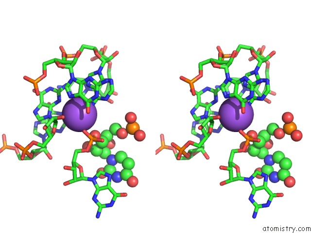
Stereo pair view
Mono view
Stereo pair view
A full contact list of Sodium with other atoms in the Na binding
site number 1 of Crystal Structure of A Mutant of the Sarcin/Ricin Domain From Rat 28S Rrna within 5.0Å range:
|
Reference:
C.C.Correll,
J.Beneken,
M.J.Plantinga,
M.Lubbers,
Y.L.Chan.
The Common and Distinctive Features of the Bulged-G Motif Based on A 1.04 A Resolution Rna Structure Nucleic Acids Res. V. 31 6806 2003.
ISSN: ISSN 0305-1048
PubMed: 14627814
DOI: 10.1093/NAR/GKG908
Page generated: Sun Oct 6 21:23:59 2024
ISSN: ISSN 0305-1048
PubMed: 14627814
DOI: 10.1093/NAR/GKG908
Last articles
Fe in 2YXOFe in 2YRS
Fe in 2YXC
Fe in 2YNM
Fe in 2YVJ
Fe in 2YP1
Fe in 2YU2
Fe in 2YU1
Fe in 2YQB
Fe in 2YOO