Sodium »
PDB 1m90-1nji »
1mvo »
Sodium in PDB 1mvo: Crystal Structure of the Phop Receiver Domain From Bacillus Subtilis
Protein crystallography data
The structure of Crystal Structure of the Phop Receiver Domain From Bacillus Subtilis, PDB code: 1mvo
was solved by
C.Birck,
Y.Chen,
F.M.Hulett,
J.P.Samama,
Structural Proteomicsin Europe (Spine),
with X-Ray Crystallography technique. A brief refinement statistics is given in the table below:
| Resolution Low / High (Å) | 27.00 / 1.60 |
| Space group | P 41 21 2 |
| Cell size a, b, c (Å), α, β, γ (°) | 45.704, 45.704, 134.811, 90.00, 90.00, 90.00 |
| R / Rfree (%) | 18.6 / 23.2 |
Other elements in 1mvo:
The structure of Crystal Structure of the Phop Receiver Domain From Bacillus Subtilis also contains other interesting chemical elements:
| Manganese | (Mn) | 1 atom |
Sodium Binding Sites:
The binding sites of Sodium atom in the Crystal Structure of the Phop Receiver Domain From Bacillus Subtilis
(pdb code 1mvo). This binding sites where shown within
5.0 Angstroms radius around Sodium atom.
In total 2 binding sites of Sodium where determined in the Crystal Structure of the Phop Receiver Domain From Bacillus Subtilis, PDB code: 1mvo:
Jump to Sodium binding site number: 1; 2;
In total 2 binding sites of Sodium where determined in the Crystal Structure of the Phop Receiver Domain From Bacillus Subtilis, PDB code: 1mvo:
Jump to Sodium binding site number: 1; 2;
Sodium binding site 1 out of 2 in 1mvo
Go back to
Sodium binding site 1 out
of 2 in the Crystal Structure of the Phop Receiver Domain From Bacillus Subtilis
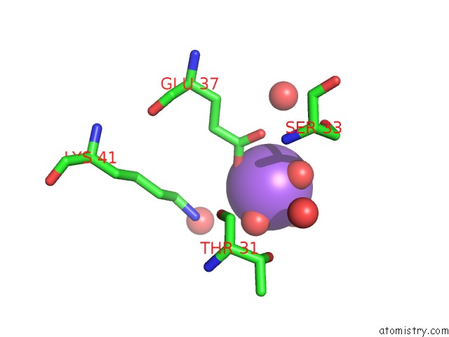
Mono view
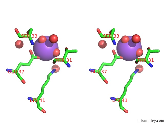
Stereo pair view
Mono view
Stereo pair view
A full contact list of Sodium with other atoms in the Na binding
site number 1 of Crystal Structure of the Phop Receiver Domain From Bacillus Subtilis within 5.0Å range:
|
Sodium binding site 2 out of 2 in 1mvo
Go back to
Sodium binding site 2 out
of 2 in the Crystal Structure of the Phop Receiver Domain From Bacillus Subtilis
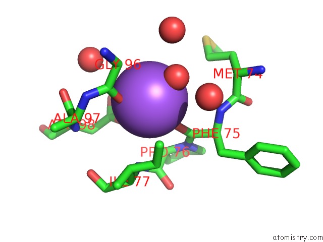
Mono view
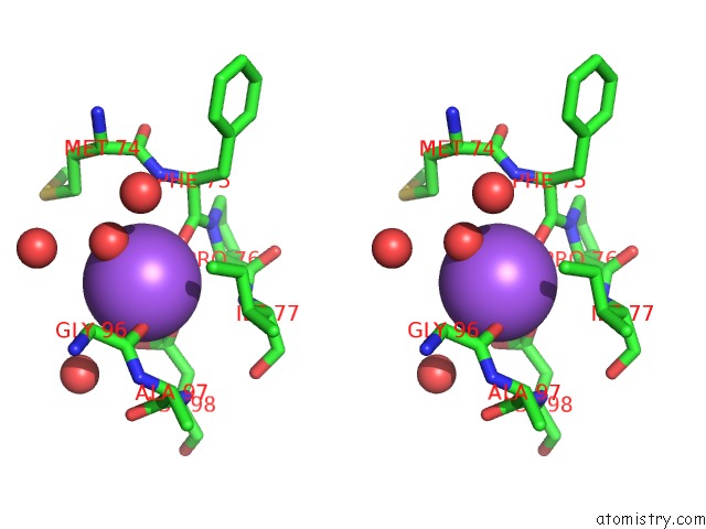
Stereo pair view
Mono view
Stereo pair view
A full contact list of Sodium with other atoms in the Na binding
site number 2 of Crystal Structure of the Phop Receiver Domain From Bacillus Subtilis within 5.0Å range:
|
Reference:
C.Birck,
Y.Chen,
F.M.Hulett,
J.P.Samama.
The Crystal Structure of the Phosphorylation Domain in Phop Reveals A Functional Tandem Association Mediated By An Asymmetric Interface J.Bacteriol. V. 185 254 2003.
ISSN: ISSN 0021-9193
PubMed: 12486062
DOI: 10.1128/JB.185.1.254-261.2003
Page generated: Sun Oct 6 20:34:37 2024
ISSN: ISSN 0021-9193
PubMed: 12486062
DOI: 10.1128/JB.185.1.254-261.2003
Last articles
K in 7NPXK in 7NWD
K in 7NXF
K in 7NF4
K in 7NHT
K in 7NKG
K in 7NF2
K in 7N9L
K in 7NFZ
K in 7NF3