Sodium »
PDB 1m90-1nji »
1mly »
Sodium in PDB 1mly: Crystal Structure of 7,8-Diaminopelargonic Acid Synthase in Complex with the Cis Isomer of Amiclenomycin
Enzymatic activity of Crystal Structure of 7,8-Diaminopelargonic Acid Synthase in Complex with the Cis Isomer of Amiclenomycin
All present enzymatic activity of Crystal Structure of 7,8-Diaminopelargonic Acid Synthase in Complex with the Cis Isomer of Amiclenomycin:
2.6.1.62;
2.6.1.62;
Protein crystallography data
The structure of Crystal Structure of 7,8-Diaminopelargonic Acid Synthase in Complex with the Cis Isomer of Amiclenomycin, PDB code: 1mly
was solved by
J.Sandmark,
S.Mann,
A.Marquet,
G.Schneider,
with X-Ray Crystallography technique. A brief refinement statistics is given in the table below:
| Resolution Low / High (Å) | 20.00 / 1.81 |
| Space group | C 1 2 1 |
| Cell size a, b, c (Å), α, β, γ (°) | 128.655, 56.073, 116.174, 90.00, 109.86, 90.00 |
| R / Rfree (%) | 19.4 / 22 |
Sodium Binding Sites:
The binding sites of Sodium atom in the Crystal Structure of 7,8-Diaminopelargonic Acid Synthase in Complex with the Cis Isomer of Amiclenomycin
(pdb code 1mly). This binding sites where shown within
5.0 Angstroms radius around Sodium atom.
In total 2 binding sites of Sodium where determined in the Crystal Structure of 7,8-Diaminopelargonic Acid Synthase in Complex with the Cis Isomer of Amiclenomycin, PDB code: 1mly:
Jump to Sodium binding site number: 1; 2;
In total 2 binding sites of Sodium where determined in the Crystal Structure of 7,8-Diaminopelargonic Acid Synthase in Complex with the Cis Isomer of Amiclenomycin, PDB code: 1mly:
Jump to Sodium binding site number: 1; 2;
Sodium binding site 1 out of 2 in 1mly
Go back to
Sodium binding site 1 out
of 2 in the Crystal Structure of 7,8-Diaminopelargonic Acid Synthase in Complex with the Cis Isomer of Amiclenomycin
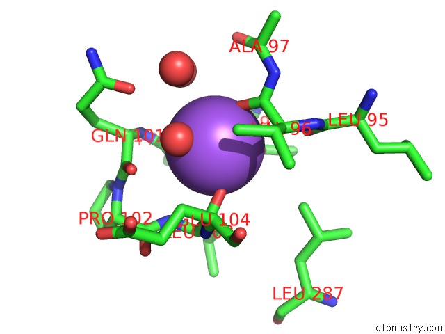
Mono view
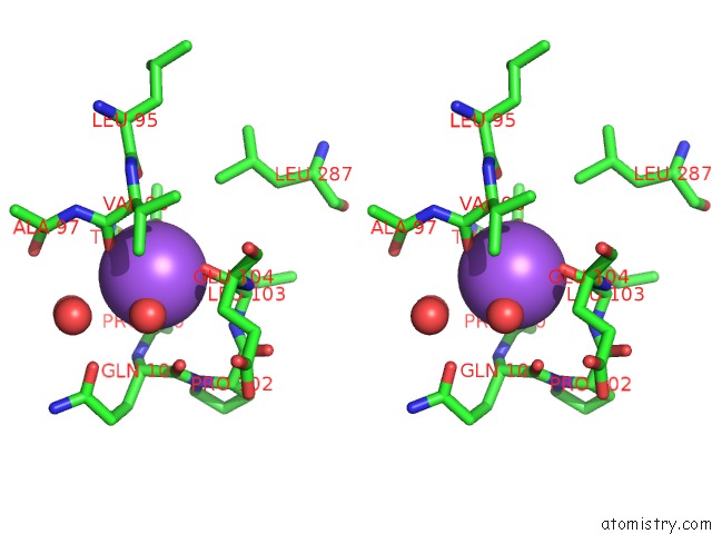
Stereo pair view
Mono view
Stereo pair view
A full contact list of Sodium with other atoms in the Na binding
site number 1 of Crystal Structure of 7,8-Diaminopelargonic Acid Synthase in Complex with the Cis Isomer of Amiclenomycin within 5.0Å range:
|
Sodium binding site 2 out of 2 in 1mly
Go back to
Sodium binding site 2 out
of 2 in the Crystal Structure of 7,8-Diaminopelargonic Acid Synthase in Complex with the Cis Isomer of Amiclenomycin
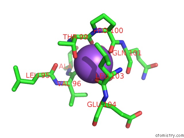
Mono view
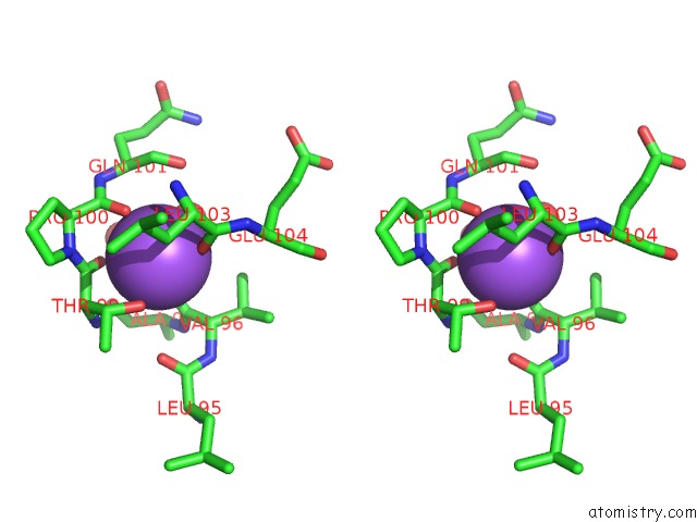
Stereo pair view
Mono view
Stereo pair view
A full contact list of Sodium with other atoms in the Na binding
site number 2 of Crystal Structure of 7,8-Diaminopelargonic Acid Synthase in Complex with the Cis Isomer of Amiclenomycin within 5.0Å range:
|
Reference:
J.Sandmark,
S.Mann,
A.Marquet,
G.Schneider.
Structural Basis For the Inhibition of the Biosynthesis of Biotin By the Antibiotic Amiclenomycin J.Biol.Chem. V. 277 43352 2002.
ISSN: ISSN 0021-9258
PubMed: 12218056
DOI: 10.1074/JBC.M207239200
Page generated: Sun Oct 6 20:32:13 2024
ISSN: ISSN 0021-9258
PubMed: 12218056
DOI: 10.1074/JBC.M207239200
Last articles
Mg in 9GUPMg in 9GUR
Mg in 9GRE
Mg in 9GTK
Mg in 9GU5
Mg in 9GOB
Mg in 9GO5
Mg in 9GMZ
Mg in 9GQO
Mg in 9GMX