Sodium »
PDB 1l0r-1m65 »
1l5b »
Sodium in PDB 1l5b: Domain-Swapped Cyanovirin-N Dimer
Protein crystallography data
The structure of Domain-Swapped Cyanovirin-N Dimer, PDB code: 1l5b
was solved by
L.G.Barrientos,
J.M.Louis,
I.Botos,
T.Mori,
Z.Han,
B.R.O'keefe,
M.R.Boyd,
A.Wlodawer,
A.M.Gronenborn,
with X-Ray Crystallography technique. A brief refinement statistics is given in the table below:
| Resolution Low / High (Å) | 19.90 / 2.00 |
| Space group | P 41 21 2 |
| Cell size a, b, c (Å), α, β, γ (°) | 61.966, 61.966, 148.400, 90.00, 90.00, 90.00 |
| R / Rfree (%) | 24.7 / 25.8 |
Sodium Binding Sites:
The binding sites of Sodium atom in the Domain-Swapped Cyanovirin-N Dimer
(pdb code 1l5b). This binding sites where shown within
5.0 Angstroms radius around Sodium atom.
In total only one binding site of Sodium was determined in the Domain-Swapped Cyanovirin-N Dimer, PDB code: 1l5b:
In total only one binding site of Sodium was determined in the Domain-Swapped Cyanovirin-N Dimer, PDB code: 1l5b:
Sodium binding site 1 out of 1 in 1l5b
Go back to
Sodium binding site 1 out
of 1 in the Domain-Swapped Cyanovirin-N Dimer
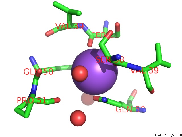
Mono view
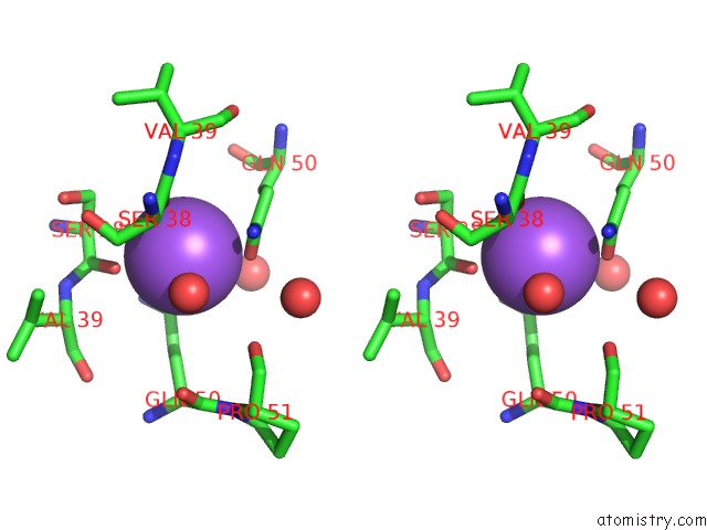
Stereo pair view
Mono view
Stereo pair view
A full contact list of Sodium with other atoms in the Na binding
site number 1 of Domain-Swapped Cyanovirin-N Dimer within 5.0Å range:
|
Reference:
L.G.Barrientos,
J.M.Louis,
I.Botos,
T.Mori,
Z.Han,
B.R.O'keefe,
M.R.Boyd,
A.Wlodawer,
A.M.Gronenborn.
The Domain-Swapped Dimer of Cyanovirin-N Is in A Metastable Folded State: Reconciliation of X-Ray and uc(Nmr) Structures. Structure V. 10 673 2002.
ISSN: ISSN 0969-2126
PubMed: 12015150
DOI: 10.1016/S0969-2126(02)00758-X
Page generated: Sun Oct 6 20:15:41 2024
ISSN: ISSN 0969-2126
PubMed: 12015150
DOI: 10.1016/S0969-2126(02)00758-X
Last articles
Mg in 1VQ4Mg in 1VPA
Mg in 1VPE
Mg in 1VOM
Mg in 1VMA
Mg in 1VMK
Mg in 1VM9
Mg in 1VCR
Mg in 1VLB
Mg in 1VKP