Sodium »
PDB 1k7e-1l0i »
1knp »
Sodium in PDB 1knp: E. Coli L-Aspartate Oxidase: Mutant R386L in Complex with Succinate
Enzymatic activity of E. Coli L-Aspartate Oxidase: Mutant R386L in Complex with Succinate
All present enzymatic activity of E. Coli L-Aspartate Oxidase: Mutant R386L in Complex with Succinate:
1.4.3.16;
1.4.3.16;
Protein crystallography data
The structure of E. Coli L-Aspartate Oxidase: Mutant R386L in Complex with Succinate, PDB code: 1knp
was solved by
R.T.Bossi,
A.Mattevi,
with X-Ray Crystallography technique. A brief refinement statistics is given in the table below:
| Resolution Low / High (Å) | 40.00 / 2.60 |
| Space group | P 41 21 2 |
| Cell size a, b, c (Å), α, β, γ (°) | 72.540, 72.540, 309.120, 90.00, 90.00, 90.00 |
| R / Rfree (%) | 23 / 28.1 |
Sodium Binding Sites:
The binding sites of Sodium atom in the E. Coli L-Aspartate Oxidase: Mutant R386L in Complex with Succinate
(pdb code 1knp). This binding sites where shown within
5.0 Angstroms radius around Sodium atom.
In total only one binding site of Sodium was determined in the E. Coli L-Aspartate Oxidase: Mutant R386L in Complex with Succinate, PDB code: 1knp:
In total only one binding site of Sodium was determined in the E. Coli L-Aspartate Oxidase: Mutant R386L in Complex with Succinate, PDB code: 1knp:
Sodium binding site 1 out of 1 in 1knp
Go back to
Sodium binding site 1 out
of 1 in the E. Coli L-Aspartate Oxidase: Mutant R386L in Complex with Succinate
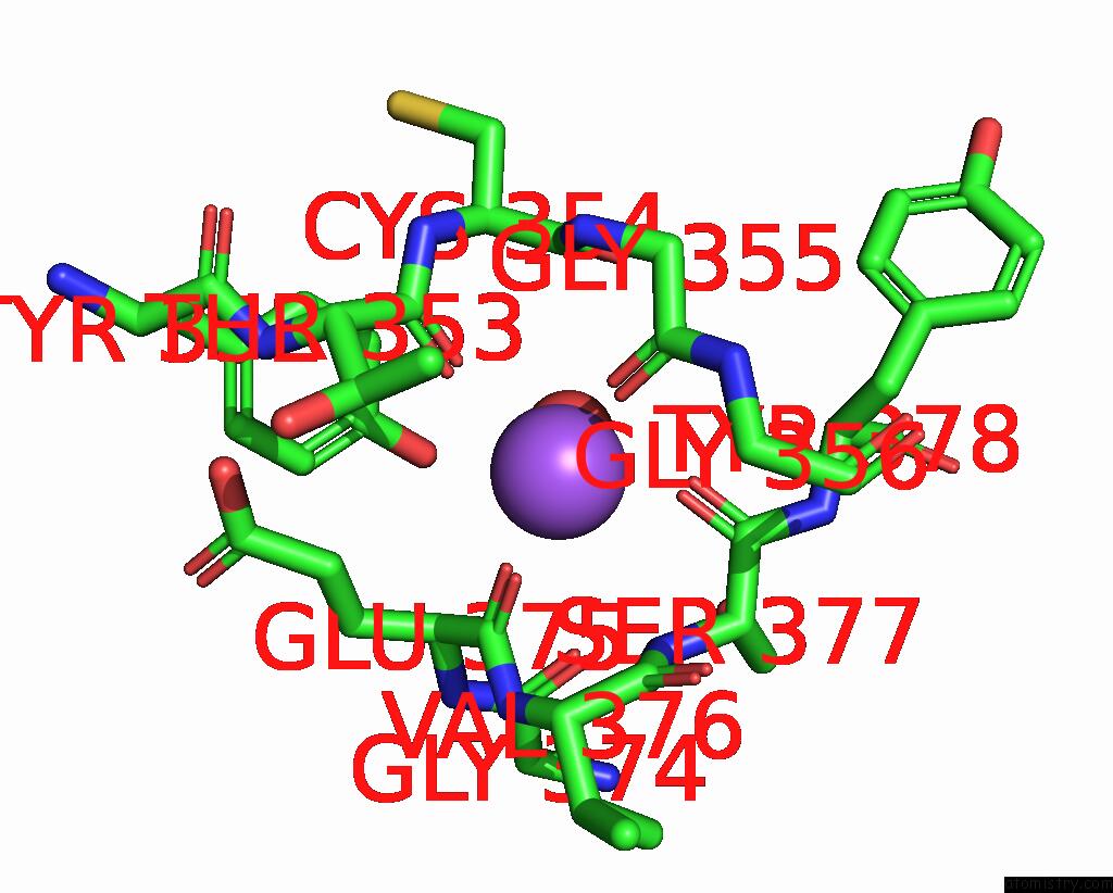
Mono view
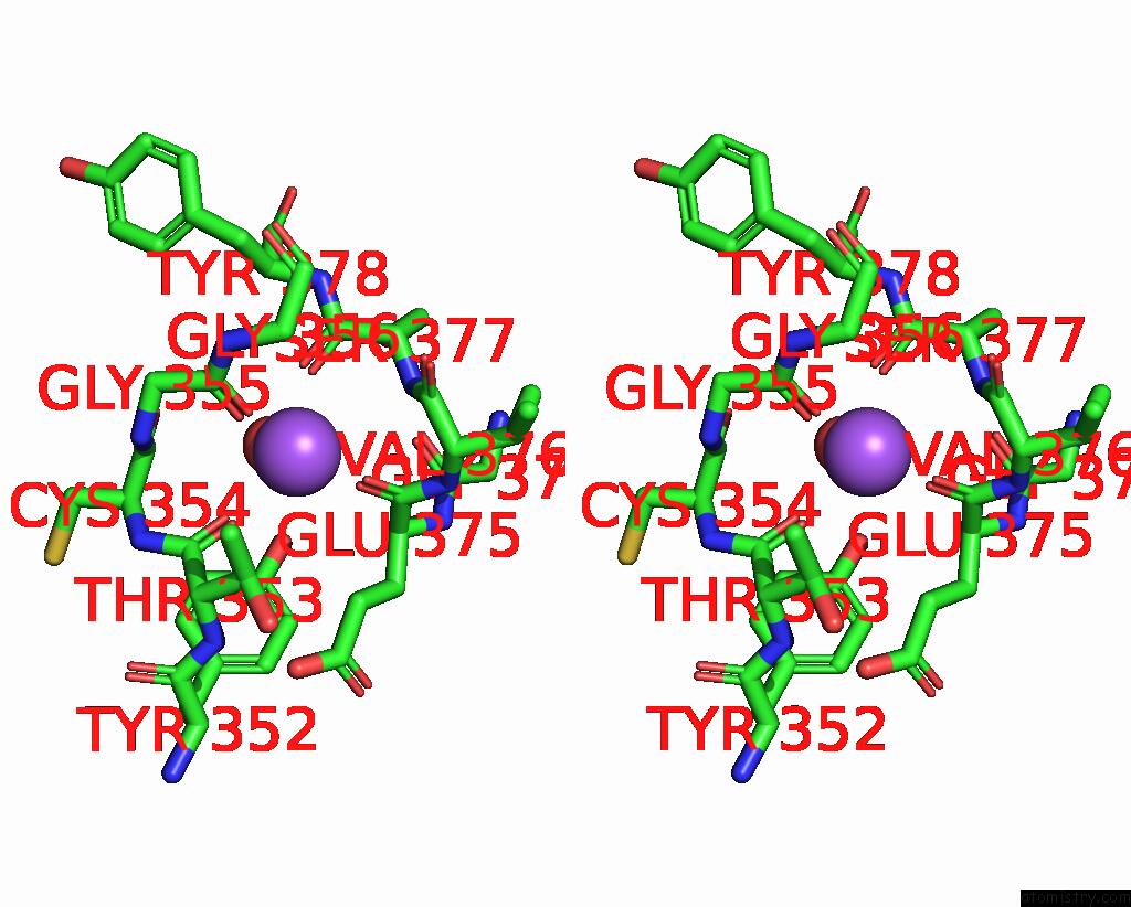
Stereo pair view
Mono view
Stereo pair view
A full contact list of Sodium with other atoms in the Na binding
site number 1 of E. Coli L-Aspartate Oxidase: Mutant R386L in Complex with Succinate within 5.0Å range:
|
Reference:
R.T.Bossi,
A.Negri,
G.Tedeschi,
A.Mattevi.
Structure of Fad-Bound L-Aspartate Oxidase: Insight Into Substrate Specificity and Catalysis. Biochemistry V. 41 3018 2002.
ISSN: ISSN 0006-2960
PubMed: 11863440
DOI: 10.1021/BI015939R
Page generated: Sun Aug 17 06:07:49 2025
ISSN: ISSN 0006-2960
PubMed: 11863440
DOI: 10.1021/BI015939R
Last articles
Zn in 3SJPZn in 3SKS
Zn in 3SJF
Zn in 3SJG
Zn in 3SJX
Zn in 3SJE
Zn in 3SEY
Zn in 3SJD
Zn in 3SJC
Zn in 3SIQ