Sodium »
PDB 1jb7-1k73 »
1jou »
Sodium in PDB 1jou: Crystal Structure of Native S195A Thrombin with An Unoccupied Active Site
Protein crystallography data
The structure of Crystal Structure of Native S195A Thrombin with An Unoccupied Active Site, PDB code: 1jou
was solved by
J.A.Huntington,
C.T.Esmon,
with X-Ray Crystallography technique. A brief refinement statistics is given in the table below:
| Resolution Low / High (Å) | 24.69 / 1.80 |
| Space group | P 1 21 1 |
| Cell size a, b, c (Å), α, β, γ (°) | 71.230, 76.580, 97.320, 90.00, 106.64, 90.00 |
| R / Rfree (%) | 22.2 / 24.5 |
Sodium Binding Sites:
The binding sites of Sodium atom in the Crystal Structure of Native S195A Thrombin with An Unoccupied Active Site
(pdb code 1jou). This binding sites where shown within
5.0 Angstroms radius around Sodium atom.
In total 3 binding sites of Sodium where determined in the Crystal Structure of Native S195A Thrombin with An Unoccupied Active Site, PDB code: 1jou:
Jump to Sodium binding site number: 1; 2; 3;
In total 3 binding sites of Sodium where determined in the Crystal Structure of Native S195A Thrombin with An Unoccupied Active Site, PDB code: 1jou:
Jump to Sodium binding site number: 1; 2; 3;
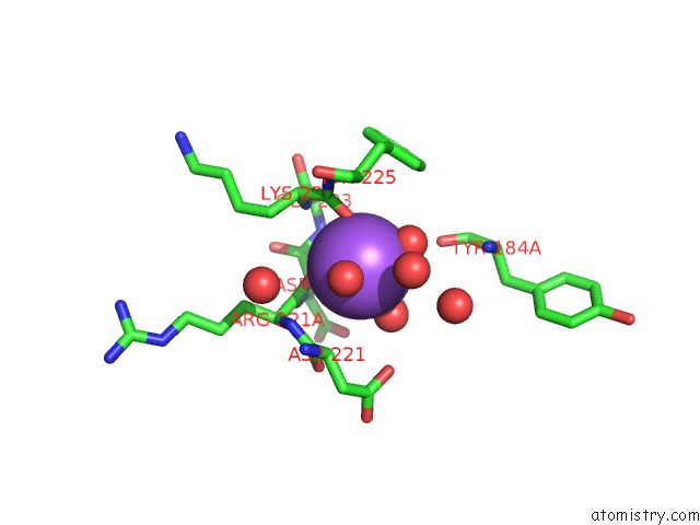
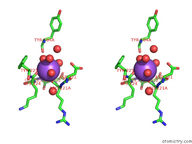
Sodium binding site 1 out of 3 in 1jou
Go back to
Sodium binding site 1 out
of 3 in the Crystal Structure of Native S195A Thrombin with An Unoccupied Active Site
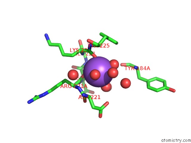
Mono view
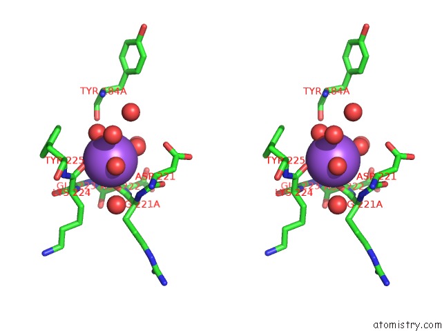
Stereo pair view
Mono view
Stereo pair view
A full contact list of Sodium with other atoms in the Na binding
site number 1 of Crystal Structure of Native S195A Thrombin with An Unoccupied Active Site within 5.0Å range:
|
Sodium binding site 2 out of 3 in 1jou
Go back to
Sodium binding site 2 out
of 3 in the Crystal Structure of Native S195A Thrombin with An Unoccupied Active Site
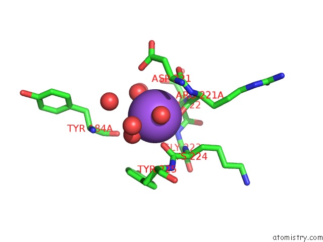
Mono view
Stereo pair view
Mono view
Stereo pair view
A full contact list of Sodium with other atoms in the Na binding
site number 2 of Crystal Structure of Native S195A Thrombin with An Unoccupied Active Site within 5.0Å range:
|
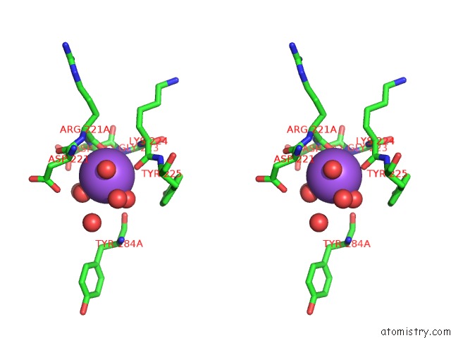
Sodium binding site 3 out of 3 in 1jou
Go back to
Sodium binding site 3 out
of 3 in the Crystal Structure of Native S195A Thrombin with An Unoccupied Active Site
Mono view
Stereo pair view
Mono view
Stereo pair view
A full contact list of Sodium with other atoms in the Na binding
site number 3 of Crystal Structure of Native S195A Thrombin with An Unoccupied Active Site within 5.0Å range:
|
Reference:
J.A.Huntington,
C.T.Esmon.
The Molecular Basis of Thrombin Allostery Revealed By A 1.8A Structure of the Slow Form Structure V. 11 469 2003.
ISSN: ISSN 0969-2126
PubMed: 12679024
DOI: 10.1016/S0969-2126(03)00049-2
Page generated: Sun Aug 17 05:21:11 2025
ISSN: ISSN 0969-2126
PubMed: 12679024
DOI: 10.1016/S0969-2126(03)00049-2
Last articles
Mn in 9LJUMn in 9LJW
Mn in 9LJS
Mn in 9LJR
Mn in 9LJT
Mn in 9LJV
Mg in 9UA2
Mg in 9R96
Mg in 9VM1
Mg in 9P01