Sodium »
PDB 1jb7-1k73 »
1jlj »
Sodium in PDB 1jlj: 1.6 Angstrom Crystal Structure of the Human Neuroreceptor Anchoring and Molybdenum Cofactor Biosynthesis Protein Gephyrin
Protein crystallography data
The structure of 1.6 Angstrom Crystal Structure of the Human Neuroreceptor Anchoring and Molybdenum Cofactor Biosynthesis Protein Gephyrin, PDB code: 1jlj
was solved by
G.Schwarz,
N.Schrader,
R.R.Mendel,
H.-J.Hecht,
H.Schindelin,
with X-Ray Crystallography technique. A brief refinement statistics is given in the table below:
| Resolution Low / High (Å) | 20.00 / 1.60 |
| Space group | C 1 2 1 |
| Cell size a, b, c (Å), α, β, γ (°) | 114.269, 66.008, 73.122, 90.00, 93.27, 90.00 |
| R / Rfree (%) | 14.7 / 18.3 |
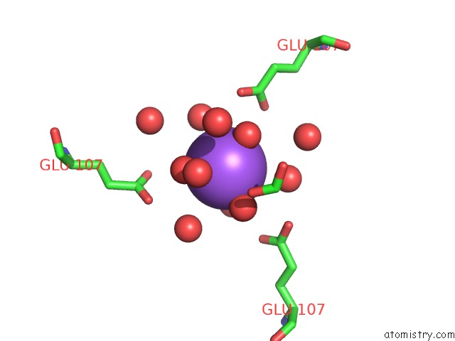
Sodium Binding Sites:
The binding sites of Sodium atom in the 1.6 Angstrom Crystal Structure of the Human Neuroreceptor Anchoring and Molybdenum Cofactor Biosynthesis Protein Gephyrin
(pdb code 1jlj). This binding sites where shown within
5.0 Angstroms radius around Sodium atom.
In total only one binding site of Sodium was determined in the 1.6 Angstrom Crystal Structure of the Human Neuroreceptor Anchoring and Molybdenum Cofactor Biosynthesis Protein Gephyrin, PDB code: 1jlj:
In total only one binding site of Sodium was determined in the 1.6 Angstrom Crystal Structure of the Human Neuroreceptor Anchoring and Molybdenum Cofactor Biosynthesis Protein Gephyrin, PDB code: 1jlj:
Sodium binding site 1 out of 1 in 1jlj
Go back to
Sodium binding site 1 out
of 1 in the 1.6 Angstrom Crystal Structure of the Human Neuroreceptor Anchoring and Molybdenum Cofactor Biosynthesis Protein Gephyrin
Mono view

Stereo pair view
Mono view
Stereo pair view
A full contact list of Sodium with other atoms in the Na binding
site number 1 of 1.6 Angstrom Crystal Structure of the Human Neuroreceptor Anchoring and Molybdenum Cofactor Biosynthesis Protein Gephyrin within 5.0Å range:
|
Reference:
G.Schwarz,
N.Schrader,
R.R.Mendel,
H.J.Hecht,
H.Schindelin.
Crystal Structures of Human Gephyrin and Plant CNX1 G Domains: Comparative Analysis and Functional Implications. J.Mol.Biol. V. 312 405 2001.
ISSN: ISSN 0022-2836
PubMed: 11554796
DOI: 10.1006/JMBI.2001.4952
Page generated: Sun Oct 6 18:56:59 2024
ISSN: ISSN 0022-2836
PubMed: 11554796
DOI: 10.1006/JMBI.2001.4952
Last articles
F in 4G2IF in 4G2H
F in 4G31
F in 4G1W
F in 4G16
F in 4FXY
F in 4FXQ
F in 4FZF
F in 4FP1
F in 4FVX