Sodium »
PDB 1ghy-1hn1 »
1gvh »
Sodium in PDB 1gvh: The X-Ray Structure of Ferric Escherichia Coli Flavohemoglobin Reveals An Unespected Geometry of the Distal Heme Pocket
Enzymatic activity of The X-Ray Structure of Ferric Escherichia Coli Flavohemoglobin Reveals An Unespected Geometry of the Distal Heme Pocket
All present enzymatic activity of The X-Ray Structure of Ferric Escherichia Coli Flavohemoglobin Reveals An Unespected Geometry of the Distal Heme Pocket:
1.6.99.7;
1.6.99.7;
Protein crystallography data
The structure of The X-Ray Structure of Ferric Escherichia Coli Flavohemoglobin Reveals An Unespected Geometry of the Distal Heme Pocket, PDB code: 1gvh
was solved by
A.Ilari,
K.A.Johnson,
A.Bonamore,
A.Farina,
A.Boffi,
with X-Ray Crystallography technique. A brief refinement statistics is given in the table below:
| Resolution Low / High (Å) | 25 / 2.19 |
| Space group | P 6 2 2 |
| Cell size a, b, c (Å), α, β, γ (°) | 164.860, 164.860, 53.460, 90.00, 90.00, 120.00 |
| R / Rfree (%) | 18.7 / 24.7 |
Other elements in 1gvh:
The structure of The X-Ray Structure of Ferric Escherichia Coli Flavohemoglobin Reveals An Unespected Geometry of the Distal Heme Pocket also contains other interesting chemical elements:
| Iron | (Fe) | 1 atom |
| Chlorine | (Cl) | 1 atom |
Sodium Binding Sites:
The binding sites of Sodium atom in the The X-Ray Structure of Ferric Escherichia Coli Flavohemoglobin Reveals An Unespected Geometry of the Distal Heme Pocket
(pdb code 1gvh). This binding sites where shown within
5.0 Angstroms radius around Sodium atom.
In total 2 binding sites of Sodium where determined in the The X-Ray Structure of Ferric Escherichia Coli Flavohemoglobin Reveals An Unespected Geometry of the Distal Heme Pocket, PDB code: 1gvh:
Jump to Sodium binding site number: 1; 2;
In total 2 binding sites of Sodium where determined in the The X-Ray Structure of Ferric Escherichia Coli Flavohemoglobin Reveals An Unespected Geometry of the Distal Heme Pocket, PDB code: 1gvh:
Jump to Sodium binding site number: 1; 2;
Sodium binding site 1 out of 2 in 1gvh
Go back to
Sodium binding site 1 out
of 2 in the The X-Ray Structure of Ferric Escherichia Coli Flavohemoglobin Reveals An Unespected Geometry of the Distal Heme Pocket
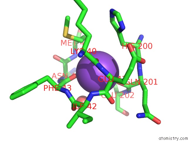
Mono view
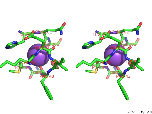
Stereo pair view
Mono view
Stereo pair view
A full contact list of Sodium with other atoms in the Na binding
site number 1 of The X-Ray Structure of Ferric Escherichia Coli Flavohemoglobin Reveals An Unespected Geometry of the Distal Heme Pocket within 5.0Å range:
|
Sodium binding site 2 out of 2 in 1gvh
Go back to
Sodium binding site 2 out
of 2 in the The X-Ray Structure of Ferric Escherichia Coli Flavohemoglobin Reveals An Unespected Geometry of the Distal Heme Pocket
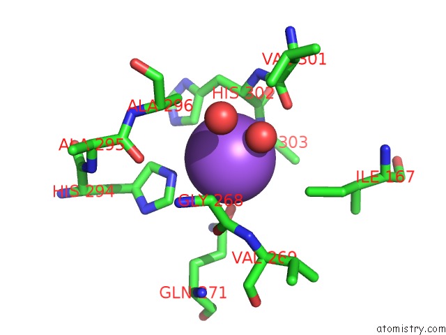
Mono view
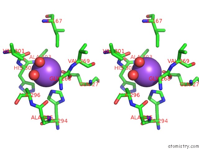
Stereo pair view
Mono view
Stereo pair view
A full contact list of Sodium with other atoms in the Na binding
site number 2 of The X-Ray Structure of Ferric Escherichia Coli Flavohemoglobin Reveals An Unespected Geometry of the Distal Heme Pocket within 5.0Å range:
|
Reference:
A.Ilari,
A.Bonamore,
A.Farina,
K.A.Johnson,
A.Boffi.
The X-Ray Structure of Ferric Escherichia Coli Flavohemoglobin Reveals An Unexpected Geometry of the Distal Heme Pocket J.Biol.Chem. V. 277 23725 2002.
ISSN: ISSN 0021-9258
PubMed: 11964402
DOI: 10.1074/JBC.M202228200
Page generated: Sun Aug 17 05:07:22 2025
ISSN: ISSN 0021-9258
PubMed: 11964402
DOI: 10.1074/JBC.M202228200
Last articles
Na in 7E4CNa in 7E21
Na in 7E1Z
Na in 7E4A
Na in 7E02
Na in 7DTF
Na in 7DTB
Na in 7DPH
Na in 7DPD
Na in 7DNB