Sodium »
PDB 1evr-1gb5 »
1exc »
Sodium in PDB 1exc: Crystal Structure of B. Subtilis Maf Protein Complexed with D-(Utp)
Protein crystallography data
The structure of Crystal Structure of B. Subtilis Maf Protein Complexed with D-(Utp), PDB code: 1exc
was solved by
G.Minasov,
M.Teplova,
G.C.Stewart,
E.V.Koonin,
W.F.Anderson,
M.Egli,
Midwest Center For Structural Genomics (Mcsg),
with X-Ray Crystallography technique. A brief refinement statistics is given in the table below:
| Resolution Low / High (Å) | 17.00 / 2.70 |
| Space group | P 21 21 21 |
| Cell size a, b, c (Å), α, β, γ (°) | 62.100, 86.580, 93.730, 90.00, 90.00, 90.00 |
| R / Rfree (%) | 19.7 / 26 |
Sodium Binding Sites:
The binding sites of Sodium atom in the Crystal Structure of B. Subtilis Maf Protein Complexed with D-(Utp)
(pdb code 1exc). This binding sites where shown within
5.0 Angstroms radius around Sodium atom.
In total only one binding site of Sodium was determined in the Crystal Structure of B. Subtilis Maf Protein Complexed with D-(Utp), PDB code: 1exc:
In total only one binding site of Sodium was determined in the Crystal Structure of B. Subtilis Maf Protein Complexed with D-(Utp), PDB code: 1exc:
Sodium binding site 1 out of 1 in 1exc
Go back to
Sodium binding site 1 out
of 1 in the Crystal Structure of B. Subtilis Maf Protein Complexed with D-(Utp)
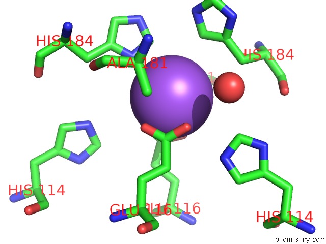
Mono view
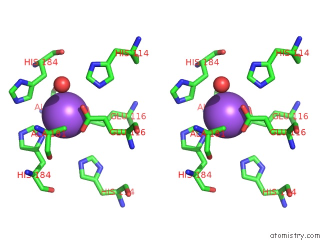
Stereo pair view
Mono view
Stereo pair view
A full contact list of Sodium with other atoms in the Na binding
site number 1 of Crystal Structure of B. Subtilis Maf Protein Complexed with D-(Utp) within 5.0Å range:
|
Reference:
G.Minasov,
M.Teplova,
G.C.Stewart,
E.V.Koonin,
W.F.Anderson,
M.Egli.
Functional Implications From Crystal Structures of the Conserved Bacillus Subtilis Protein Maf with and Without Dutp. Proc.Natl.Acad.Sci.Usa V. 97 6328 2000.
ISSN: ISSN 0027-8424
PubMed: 10841541
DOI: 10.1073/PNAS.97.12.6328
Page generated: Sun Aug 17 04:59:37 2025
ISSN: ISSN 0027-8424
PubMed: 10841541
DOI: 10.1073/PNAS.97.12.6328
Last articles
Na in 2BIINa in 2BIV
Na in 2BJB
Na in 2BFG
Na in 2BHP
Na in 2BHC
Na in 2BER
Na in 2BGD
Na in 2BDR
Na in 2BCV