Sodium »
PDB 8pkw-8qay »
8pof »
Sodium in PDB 8pof: The Crystal Structure of RSSYMEG1 Reveals A Unique Form of Smaller GH7 Endoglucanases Alongside GH7 Cellobiohydrolases in Protist Symbionts of Termites
Protein crystallography data
The structure of The Crystal Structure of RSSYMEG1 Reveals A Unique Form of Smaller GH7 Endoglucanases Alongside GH7 Cellobiohydrolases in Protist Symbionts of Termites, PDB code: 8pof
was solved by
T.Haataja,
M.Sandgren,
H.Hansson,
J.Stahlberg,
with X-Ray Crystallography technique. A brief refinement statistics is given in the table below:
| Resolution Low / High (Å) | 45.80 / 1.85 |
| Space group | I 41 |
| Cell size a, b, c (Å), α, β, γ (°) | 114.604, 114.604, 49.963, 90, 90, 90 |
| R / Rfree (%) | 16.6 / 18.7 |
Sodium Binding Sites:
The binding sites of Sodium atom in the The Crystal Structure of RSSYMEG1 Reveals A Unique Form of Smaller GH7 Endoglucanases Alongside GH7 Cellobiohydrolases in Protist Symbionts of Termites
(pdb code 8pof). This binding sites where shown within
5.0 Angstroms radius around Sodium atom.
In total only one binding site of Sodium was determined in the The Crystal Structure of RSSYMEG1 Reveals A Unique Form of Smaller GH7 Endoglucanases Alongside GH7 Cellobiohydrolases in Protist Symbionts of Termites, PDB code: 8pof:
In total only one binding site of Sodium was determined in the The Crystal Structure of RSSYMEG1 Reveals A Unique Form of Smaller GH7 Endoglucanases Alongside GH7 Cellobiohydrolases in Protist Symbionts of Termites, PDB code: 8pof:
Sodium binding site 1 out of 1 in 8pof
Go back to
Sodium binding site 1 out
of 1 in the The Crystal Structure of RSSYMEG1 Reveals A Unique Form of Smaller GH7 Endoglucanases Alongside GH7 Cellobiohydrolases in Protist Symbionts of Termites
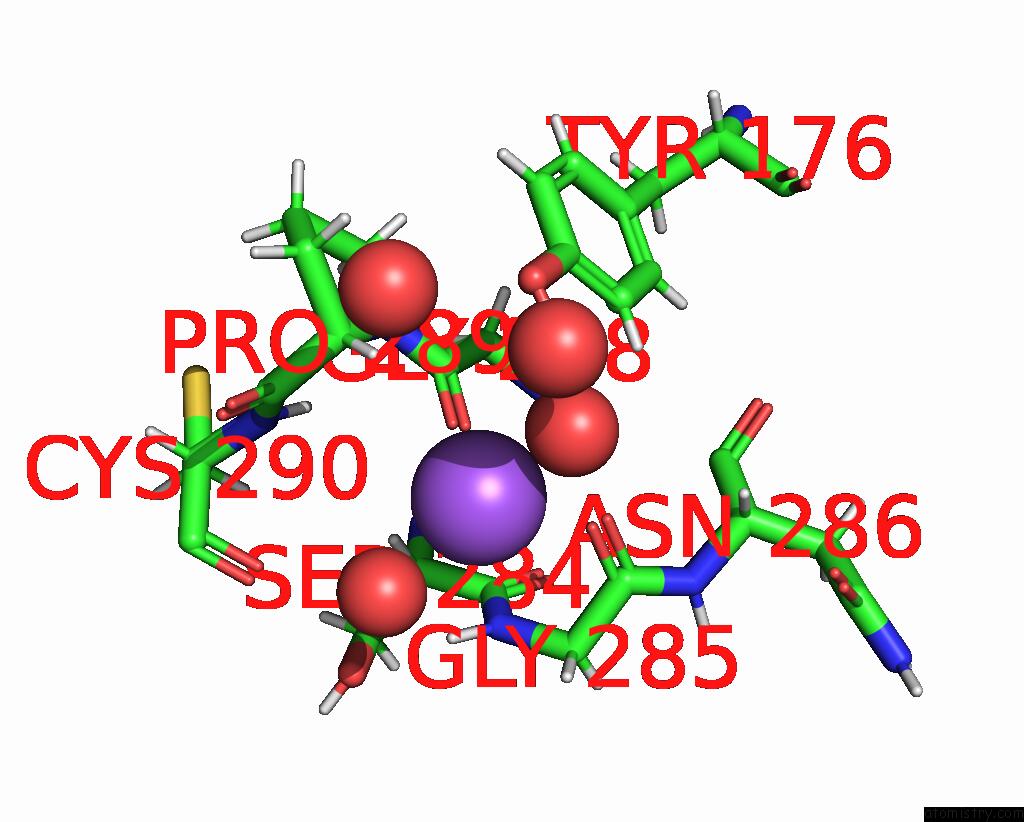
Mono view
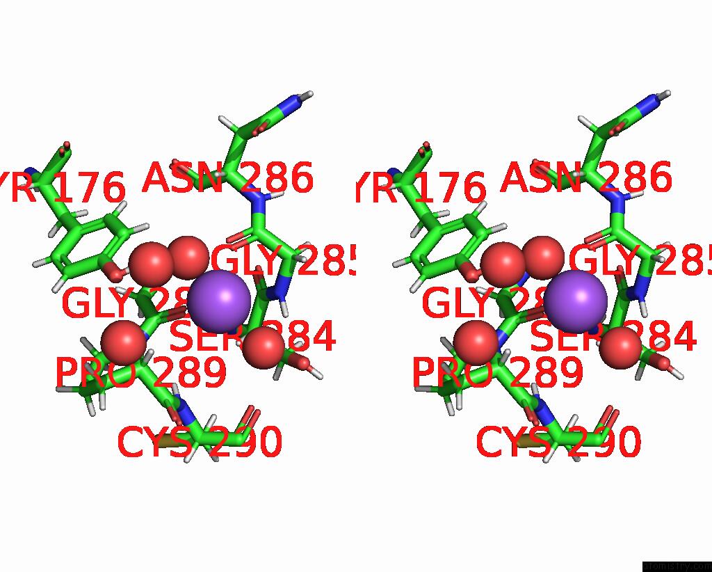
Stereo pair view
Mono view
Stereo pair view
A full contact list of Sodium with other atoms in the Na binding
site number 1 of The Crystal Structure of RSSYMEG1 Reveals A Unique Form of Smaller GH7 Endoglucanases Alongside GH7 Cellobiohydrolases in Protist Symbionts of Termites within 5.0Å range:
|
Reference:
T.Haataja,
H.Hansson,
S.Moriya,
M.Sandgren,
J.Stahlberg.
The Crystal Structure of RSSYMEG1 Reveals A Unique Form of Smaller GH7 Endoglucanases Alongside GH7 Cellobiohydrolases in Protist Symbionts of Termites. Febs J. 2023.
ISSN: ISSN 1742-464X
PubMed: 38073120
DOI: 10.1111/FEBS.17029
Page generated: Mon Aug 18 15:05:43 2025
ISSN: ISSN 1742-464X
PubMed: 38073120
DOI: 10.1111/FEBS.17029
Last articles
Ni in 3ZEZNi in 3X2Z
Ni in 3ZEA
Ni in 3ZE9
Ni in 3ZE8
Ni in 3WD7
Ni in 3ZE7
Ni in 3X2Y
Ni in 3ZE6
Ni in 3X30