Sodium in PDB 7s9j: Crystal Structure of Dna Polymerase Beta with Fapy-Dg Base-Paired with A Dc
Enzymatic activity of Crystal Structure of Dna Polymerase Beta with Fapy-Dg Base-Paired with A Dc
All present enzymatic activity of Crystal Structure of Dna Polymerase Beta with Fapy-Dg Base-Paired with A Dc:
2.7.7.7;
Protein crystallography data
The structure of Crystal Structure of Dna Polymerase Beta with Fapy-Dg Base-Paired with A Dc, PDB code: 7s9j
was solved by
B.D.Freudenthal,
B.J.Ryan,
with X-Ray Crystallography technique. A brief refinement statistics is given in the table below:
Resolution Low / High (Å)
|
24.86 /
1.91
|
Space group
|
P 1 21 1
|
Cell size a, b, c (Å), α, β, γ (°)
|
54.383,
79.494,
54.885,
90,
105.72,
90
|
R / Rfree (%)
|
18.8 /
23.8
|
Sodium Binding Sites:
The binding sites of Sodium atom in the Crystal Structure of Dna Polymerase Beta with Fapy-Dg Base-Paired with A Dc
(pdb code 7s9j). This binding sites where shown within
5.0 Angstroms radius around Sodium atom.
In total 4 binding sites of Sodium where determined in the
Crystal Structure of Dna Polymerase Beta with Fapy-Dg Base-Paired with A Dc, PDB code: 7s9j:
Jump to Sodium binding site number:
1;
2;
3;
4;
Sodium binding site 1 out
of 4 in 7s9j
Go back to
Sodium Binding Sites List in 7s9j
Sodium binding site 1 out
of 4 in the Crystal Structure of Dna Polymerase Beta with Fapy-Dg Base-Paired with A Dc
Mono view
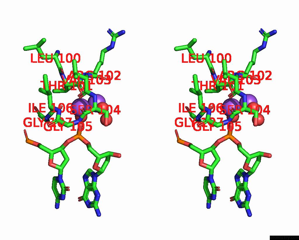
Stereo pair view
|
A full contact list of Sodium with other atoms in the Na binding
site number 1 of Crystal Structure of Dna Polymerase Beta with Fapy-Dg Base-Paired with A Dc within 5.0Å range:
probe
|
atom
|
residue
|
distance (Å)
|
B
|
Occ
|
A:Na401
b:14.2
occ:1.00
|
O
|
A:THR101
|
2.3
|
13.6
|
1.0
|
OP1
|
P:DG9
|
2.3
|
10.9
|
1.0
|
O
|
A:VAL103
|
2.4
|
10.8
|
1.0
|
O
|
A:ILE106
|
2.4
|
11.7
|
1.0
|
O
|
P:HOH115
|
2.6
|
12.3
|
1.0
|
O
|
A:HOH769
|
2.7
|
15.1
|
1.0
|
C
|
A:VAL103
|
3.3
|
15.5
|
1.0
|
C
|
A:THR101
|
3.3
|
14.3
|
1.0
|
NA
|
A:NA403
|
3.3
|
27.8
|
1.0
|
C
|
A:ILE106
|
3.4
|
9.5
|
1.0
|
P
|
P:DG9
|
3.5
|
13.8
|
1.0
|
N
|
A:ILE106
|
3.7
|
12.0
|
1.0
|
OP2
|
P:DG9
|
3.7
|
12.9
|
1.0
|
CA
|
A:SER104
|
3.8
|
13.1
|
1.0
|
CA
|
A:THR101
|
3.9
|
11.7
|
1.0
|
N
|
A:SER104
|
3.9
|
13.2
|
1.0
|
N
|
A:GLY105
|
4.0
|
11.7
|
1.0
|
CA
|
A:ILE106
|
4.0
|
11.3
|
1.0
|
N
|
A:VAL103
|
4.2
|
11.9
|
1.0
|
C
|
A:SER104
|
4.3
|
9.4
|
1.0
|
C
|
A:ARG102
|
4.3
|
17.9
|
1.0
|
N
|
A:GLY107
|
4.3
|
9.1
|
1.0
|
N
|
A:ARG102
|
4.4
|
14.5
|
1.0
|
CA
|
A:VAL103
|
4.4
|
13.9
|
1.0
|
CB
|
A:THR101
|
4.4
|
14.7
|
1.0
|
CB
|
A:ILE106
|
4.4
|
14.2
|
1.0
|
O3'
|
P:DC8
|
4.5
|
12.9
|
1.0
|
C
|
A:GLY105
|
4.6
|
10.9
|
1.0
|
CA
|
A:ARG102
|
4.6
|
15.3
|
1.0
|
O5'
|
P:DG9
|
4.6
|
13.3
|
1.0
|
CA
|
A:GLY107
|
4.7
|
12.0
|
1.0
|
O
|
A:ARG102
|
4.7
|
14.1
|
1.0
|
CA
|
A:GLY105
|
4.8
|
12.7
|
1.0
|
O
|
A:HOH790
|
4.9
|
29.8
|
1.0
|
O
|
A:LEU100
|
4.9
|
15.2
|
1.0
|
|
Sodium binding site 2 out
of 4 in 7s9j
Go back to
Sodium Binding Sites List in 7s9j
Sodium binding site 2 out
of 4 in the Crystal Structure of Dna Polymerase Beta with Fapy-Dg Base-Paired with A Dc
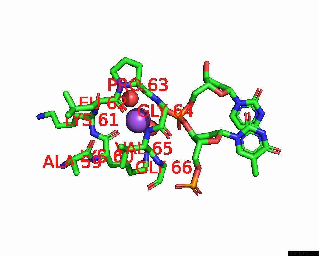
Mono view
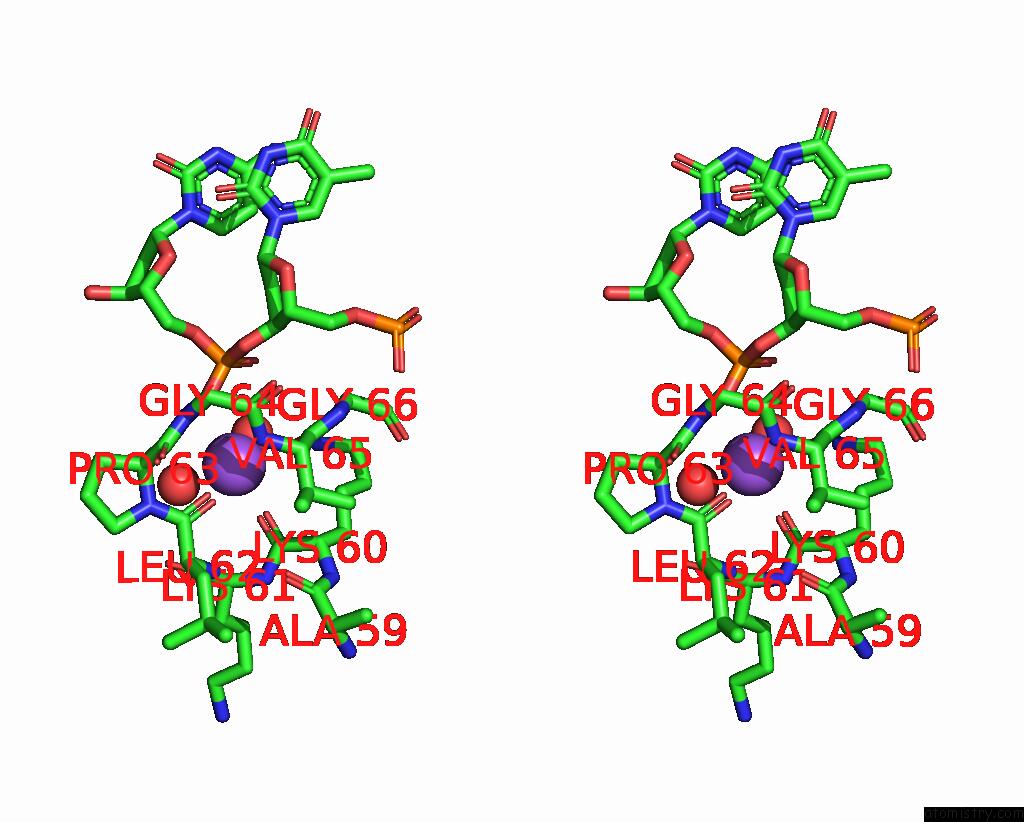
Stereo pair view
|
A full contact list of Sodium with other atoms in the Na binding
site number 2 of Crystal Structure of Dna Polymerase Beta with Fapy-Dg Base-Paired with A Dc within 5.0Å range:
probe
|
atom
|
residue
|
distance (Å)
|
B
|
Occ
|
A:Na402
b:20.3
occ:1.00
|
O
|
A:VAL65
|
2.3
|
21.6
|
1.0
|
O
|
D:HOH114
|
2.4
|
20.5
|
1.0
|
O
|
A:LEU62
|
2.4
|
22.6
|
1.0
|
O
|
A:LYS60
|
2.4
|
20.0
|
1.0
|
O
|
A:HOH725
|
2.5
|
23.5
|
1.0
|
OP1
|
D:DC3
|
2.7
|
18.4
|
1.0
|
C
|
A:VAL65
|
3.4
|
19.8
|
1.0
|
C
|
A:LEU62
|
3.4
|
24.5
|
1.0
|
C
|
A:LYS60
|
3.5
|
24.1
|
1.0
|
P
|
D:DC3
|
3.7
|
17.5
|
1.0
|
N
|
A:VAL65
|
3.8
|
23.4
|
1.0
|
OP2
|
D:DC3
|
3.9
|
18.0
|
1.0
|
CA
|
A:PRO63
|
4.1
|
25.9
|
1.0
|
N
|
A:LEU62
|
4.1
|
21.4
|
1.0
|
C
|
A:LYS61
|
4.1
|
23.7
|
1.0
|
CA
|
A:VAL65
|
4.1
|
19.7
|
1.0
|
N
|
A:GLY64
|
4.1
|
21.4
|
1.0
|
N
|
A:PRO63
|
4.2
|
22.7
|
1.0
|
CA
|
A:LYS60
|
4.2
|
19.5
|
1.0
|
O
|
A:LYS61
|
4.4
|
21.8
|
1.0
|
CA
|
A:LEU62
|
4.4
|
22.9
|
1.0
|
N
|
A:LYS61
|
4.4
|
23.8
|
1.0
|
CB
|
A:VAL65
|
4.5
|
21.6
|
1.0
|
C
|
A:PRO63
|
4.5
|
23.1
|
1.0
|
CG
|
A:LYS60
|
4.5
|
27.0
|
1.0
|
N
|
A:GLY66
|
4.5
|
15.4
|
1.0
|
CA
|
A:LYS61
|
4.5
|
22.7
|
1.0
|
O3'
|
D:DT2
|
4.6
|
18.5
|
1.0
|
CA
|
A:GLY66
|
4.7
|
17.2
|
1.0
|
C
|
A:GLY64
|
4.8
|
21.9
|
1.0
|
O
|
A:ALA59
|
4.9
|
19.3
|
1.0
|
CB
|
A:LYS60
|
5.0
|
20.0
|
1.0
|
O5'
|
D:DC3
|
5.0
|
15.5
|
1.0
|
|
Sodium binding site 3 out
of 4 in 7s9j
Go back to
Sodium Binding Sites List in 7s9j
Sodium binding site 3 out
of 4 in the Crystal Structure of Dna Polymerase Beta with Fapy-Dg Base-Paired with A Dc
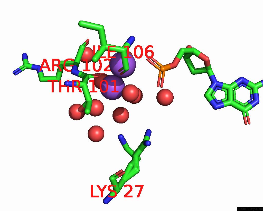
Mono view
Stereo pair view
|
A full contact list of Sodium with other atoms in the Na binding
site number 3 of Crystal Structure of Dna Polymerase Beta with Fapy-Dg Base-Paired with A Dc within 5.0Å range:
probe
|
atom
|
residue
|
distance (Å)
|
B
|
Occ
|
A:Na403
b:27.8
occ:1.00
|
O
|
A:HOH809
|
2.4
|
18.9
|
1.0
|
O
|
A:HOH545
|
2.4
|
17.9
|
1.0
|
O
|
A:THR101
|
2.5
|
13.6
|
1.0
|
O
|
A:HOH790
|
2.5
|
29.8
|
1.0
|
O
|
P:HOH115
|
2.6
|
12.3
|
1.0
|
O
|
A:HOH769
|
2.8
|
15.1
|
1.0
|
O
|
A:HOH568
|
3.2
|
36.5
|
1.0
|
NA
|
A:NA401
|
3.3
|
14.2
|
1.0
|
C
|
A:THR101
|
3.4
|
14.3
|
1.0
|
CA
|
A:ARG102
|
3.9
|
15.3
|
1.0
|
N
|
A:ARG102
|
4.1
|
14.5
|
1.0
|
OG1
|
A:THR101
|
4.3
|
12.8
|
1.0
|
CB
|
A:THR101
|
4.3
|
14.7
|
1.0
|
O
|
A:HOH813
|
4.5
|
26.9
|
1.0
|
CA
|
A:THR101
|
4.5
|
11.7
|
1.0
|
C
|
A:ARG102
|
4.5
|
17.9
|
1.0
|
NZ
|
A:LYS27
|
4.6
|
19.5
|
0.5
|
O
|
A:ARG102
|
4.7
|
14.1
|
1.0
|
O
|
A:HOH580
|
4.8
|
21.6
|
1.0
|
O
|
A:HOH635
|
4.8
|
26.4
|
1.0
|
O
|
P:HOH121
|
4.9
|
27.9
|
1.0
|
OP1
|
P:DG9
|
4.9
|
10.9
|
1.0
|
OP2
|
P:DG9
|
5.0
|
12.9
|
1.0
|
O
|
A:ILE106
|
5.0
|
11.7
|
1.0
|
|
Sodium binding site 4 out
of 4 in 7s9j
Go back to
Sodium Binding Sites List in 7s9j
Sodium binding site 4 out
of 4 in the Crystal Structure of Dna Polymerase Beta with Fapy-Dg Base-Paired with A Dc
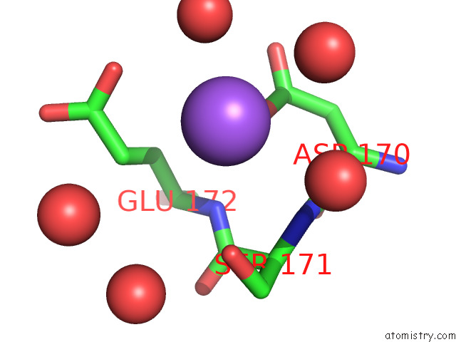
Mono view
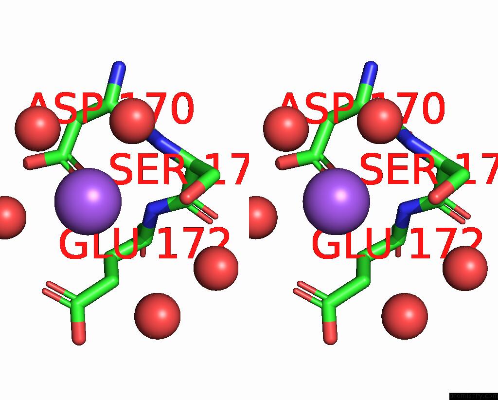
Stereo pair view
|
A full contact list of Sodium with other atoms in the Na binding
site number 4 of Crystal Structure of Dna Polymerase Beta with Fapy-Dg Base-Paired with A Dc within 5.0Å range:
probe
|
atom
|
residue
|
distance (Å)
|
B
|
Occ
|
A:Na404
b:39.0
occ:1.00
|
OD1
|
A:ASP170
|
2.8
|
18.6
|
1.0
|
O
|
A:HOH785
|
3.0
|
28.2
|
1.0
|
O
|
A:HOH726
|
3.2
|
17.8
|
1.0
|
OG
|
A:SER171
|
3.4
|
16.0
|
1.0
|
O
|
A:HOH534
|
3.6
|
16.4
|
1.0
|
CG
|
A:ASP170
|
3.7
|
17.2
|
1.0
|
N
|
A:SER171
|
3.9
|
13.7
|
1.0
|
CG
|
A:GLU172
|
4.0
|
21.1
|
1.0
|
OE1
|
A:GLU172
|
4.1
|
37.5
|
1.0
|
N
|
A:GLU172
|
4.1
|
13.6
|
1.0
|
OD2
|
A:ASP170
|
4.2
|
18.9
|
1.0
|
CD
|
A:GLU172
|
4.4
|
36.9
|
1.0
|
CB
|
A:GLU172
|
4.5
|
20.4
|
1.0
|
CB
|
A:SER171
|
4.5
|
14.5
|
1.0
|
O
|
A:HOH812
|
4.6
|
23.1
|
1.0
|
C
|
A:ASP170
|
4.6
|
16.3
|
1.0
|
CA
|
A:SER171
|
4.6
|
15.9
|
1.0
|
CA
|
A:ASP170
|
4.6
|
13.3
|
1.0
|
C
|
A:SER171
|
4.7
|
19.8
|
1.0
|
CB
|
A:ASP170
|
4.8
|
14.2
|
1.0
|
CA
|
A:GLU172
|
4.9
|
22.6
|
1.0
|
O
|
A:HOH566
|
5.0
|
19.7
|
1.0
|
|
Reference:
B.J.Ryan,
H.Yang,
J.H.T.Bacurio,
M.R.Smith,
A.K.Basu,
M.M.Greenberg,
B.D.Freudenthal.
Structural Dynamics of A Common Mutagenic Oxidative Dna Lesion in Duplex Dna and During Dna Replication. J.Am.Chem.Soc. V. 144 8054 2022.
ISSN: ESSN 1520-5126
PubMed: 35499923
DOI: 10.1021/JACS.2C00193
Page generated: Wed Oct 9 08:54:35 2024
|