Sodium »
PDB 7bqj-7cdu »
7c75 »
Sodium in PDB 7c75: Crystal Structure of Yak Lactoperoxidase with Partially Coordinated Na Ion in the Distal Heme Cavity
Protein crystallography data
The structure of Crystal Structure of Yak Lactoperoxidase with Partially Coordinated Na Ion in the Distal Heme Cavity, PDB code: 7c75
was solved by
P.K.Singh,
V.Viswanathan,
C.Rani,
N.Ahmad,
P.Sharma,
P.Kaur,
S.Sharma,
T.P.Singh,
with X-Ray Crystallography technique. A brief refinement statistics is given in the table below:
| Resolution Low / High (Å) | 64.30 / 2.70 |
| Space group | P 21 21 21 |
| Cell size a, b, c (Å), α, β, γ (°) | 79.910, 84.830, 98.560, 90.00, 90.00, 90.00 |
| R / Rfree (%) | 19.3 / 26.8 |
Other elements in 7c75:
The structure of Crystal Structure of Yak Lactoperoxidase with Partially Coordinated Na Ion in the Distal Heme Cavity also contains other interesting chemical elements:
| Potassium | (K) | 1 atom |
| Zinc | (Zn) | 1 atom |
| Iron | (Fe) | 1 atom |
| Calcium | (Ca) | 1 atom |
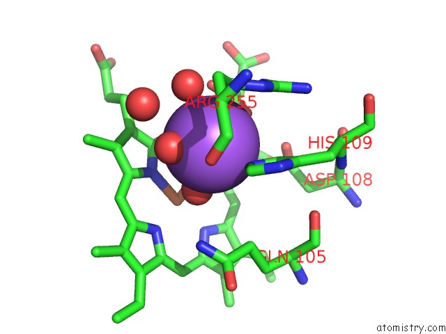
Sodium Binding Sites:
The binding sites of Sodium atom in the Crystal Structure of Yak Lactoperoxidase with Partially Coordinated Na Ion in the Distal Heme Cavity
(pdb code 7c75). This binding sites where shown within
5.0 Angstroms radius around Sodium atom.
In total only one binding site of Sodium was determined in the Crystal Structure of Yak Lactoperoxidase with Partially Coordinated Na Ion in the Distal Heme Cavity, PDB code: 7c75:
In total only one binding site of Sodium was determined in the Crystal Structure of Yak Lactoperoxidase with Partially Coordinated Na Ion in the Distal Heme Cavity, PDB code: 7c75:
Sodium binding site 1 out of 1 in 7c75
Go back to
Sodium binding site 1 out
of 1 in the Crystal Structure of Yak Lactoperoxidase with Partially Coordinated Na Ion in the Distal Heme Cavity
Mono view
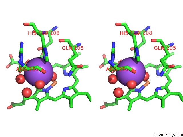
Stereo pair view
Mono view
Stereo pair view
A full contact list of Sodium with other atoms in the Na binding
site number 1 of Crystal Structure of Yak Lactoperoxidase with Partially Coordinated Na Ion in the Distal Heme Cavity within 5.0Å range:
|
Reference:
P.K.Singh,
V.Viswanathan,
C.Rani,
N.Ahmad,
P.Sharma,
P.Kaur,
S.Sharma,
T.P.Singh.
Crystal Structure of Yak Lactoperoxidase with Partially Coordinated Na Ion in the Distal Heme Cavity To Be Published.
Page generated: Tue Oct 8 16:19:37 2024
Last articles
Cl in 7VP8Cl in 7VOE
Cl in 7VOD
Cl in 7VKH
Cl in 7VOB
Cl in 7VMG
Cl in 7VO7
Cl in 7VMJ
Cl in 7VKA
Cl in 7VKG