Sodium »
PDB 7b7h-7bov »
7bgg »
Sodium in PDB 7bgg: Crystal Structure of the Heterocyclic Toxin Methyltransferase From Mycobacterium Tuberculosis
Enzymatic activity of Crystal Structure of the Heterocyclic Toxin Methyltransferase From Mycobacterium Tuberculosis
All present enzymatic activity of Crystal Structure of the Heterocyclic Toxin Methyltransferase From Mycobacterium Tuberculosis:
2.1.1.374;
2.1.1.374;
Protein crystallography data
The structure of Crystal Structure of the Heterocyclic Toxin Methyltransferase From Mycobacterium Tuberculosis, PDB code: 7bgg
was solved by
L.Denkhaus,
P.Sartor,
O.Einsle,
S.Gerhardt,
S.Fetzner,
with X-Ray Crystallography technique. A brief refinement statistics is given in the table below:
| Resolution Low / High (Å) | 15.02 / 1.04 |
| Space group | P 31 2 1 |
| Cell size a, b, c (Å), α, β, γ (°) | 70.235, 70.235, 96.449, 90, 90, 120 |
| R / Rfree (%) | 17.5 / 18.1 |
Sodium Binding Sites:
The binding sites of Sodium atom in the Crystal Structure of the Heterocyclic Toxin Methyltransferase From Mycobacterium Tuberculosis
(pdb code 7bgg). This binding sites where shown within
5.0 Angstroms radius around Sodium atom.
In total 2 binding sites of Sodium where determined in the Crystal Structure of the Heterocyclic Toxin Methyltransferase From Mycobacterium Tuberculosis, PDB code: 7bgg:
Jump to Sodium binding site number: 1; 2;
In total 2 binding sites of Sodium where determined in the Crystal Structure of the Heterocyclic Toxin Methyltransferase From Mycobacterium Tuberculosis, PDB code: 7bgg:
Jump to Sodium binding site number: 1; 2;
Sodium binding site 1 out of 2 in 7bgg
Go back to
Sodium binding site 1 out
of 2 in the Crystal Structure of the Heterocyclic Toxin Methyltransferase From Mycobacterium Tuberculosis
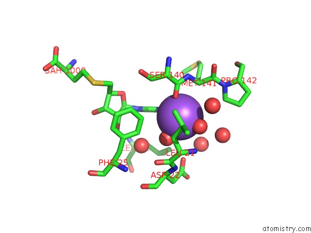
Mono view
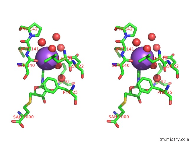
Stereo pair view
Mono view
Stereo pair view
A full contact list of Sodium with other atoms in the Na binding
site number 1 of Crystal Structure of the Heterocyclic Toxin Methyltransferase From Mycobacterium Tuberculosis within 5.0Å range:
|
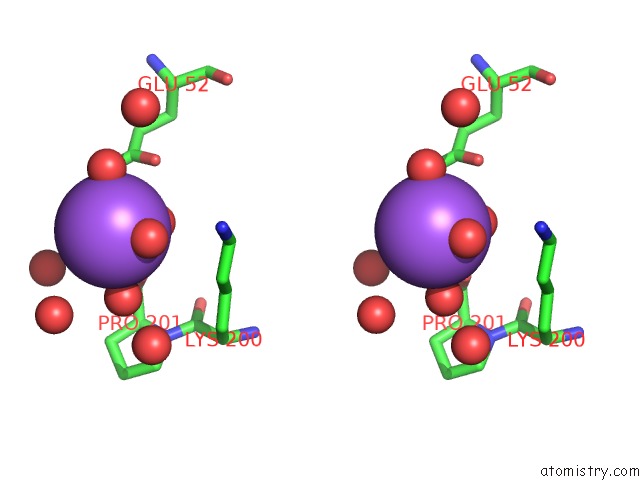
Sodium binding site 2 out of 2 in 7bgg
Go back to
Sodium binding site 2 out
of 2 in the Crystal Structure of the Heterocyclic Toxin Methyltransferase From Mycobacterium Tuberculosis
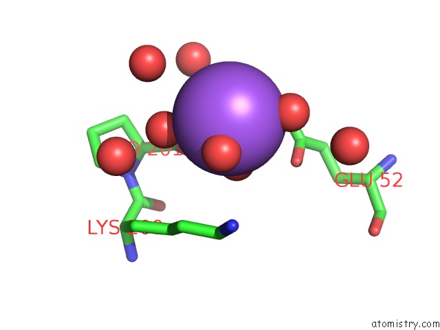
Mono view
Stereo pair view
Mono view
Stereo pair view
A full contact list of Sodium with other atoms in the Na binding
site number 2 of Crystal Structure of the Heterocyclic Toxin Methyltransferase From Mycobacterium Tuberculosis within 5.0Å range:
|
Reference:
P.Sartor,
L.Denkhaus,
S.Gerhardt,
O.Einsle,
S.Fetzner.
Structural Basis of O-Methylation of (2-Heptyl-)1-Hydroxyquinolin-4(1H)-One and Related Compounds By the Heterocyclic Toxin Methyltransferase RV0560C of Mycobacterium Tuberculosis J.Struct.Biol. 07794 2021.
ISSN: ESSN 1095-8657
DOI: 10.1016/J.JSB.2021.107794
Page generated: Tue Oct 8 16:12:34 2024
ISSN: ESSN 1095-8657
DOI: 10.1016/J.JSB.2021.107794
Last articles
Zn in 9J0NZn in 9J0O
Zn in 9J0P
Zn in 9FJX
Zn in 9EKB
Zn in 9C0F
Zn in 9CAH
Zn in 9CH0
Zn in 9CH3
Zn in 9CH1