Sodium »
PDB 7a33-7ap3 »
7a6a »
Sodium in PDB 7a6a: 1.15 A Structure of Human Apoferritin Obtained From Titan Mono- Bcor Microscope
Enzymatic activity of 1.15 A Structure of Human Apoferritin Obtained From Titan Mono- Bcor Microscope
All present enzymatic activity of 1.15 A Structure of Human Apoferritin Obtained From Titan Mono- Bcor Microscope:
1.16.3.1;
1.16.3.1;
Sodium Binding Sites:
Pages:
>>> Page 1 <<< Page 2, Binding sites: 11 - 20; Page 3, Binding sites: 21 - 30; Page 4, Binding sites: 31 - 32;Binding sites:
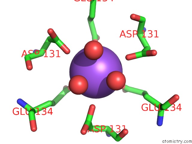
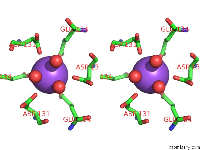
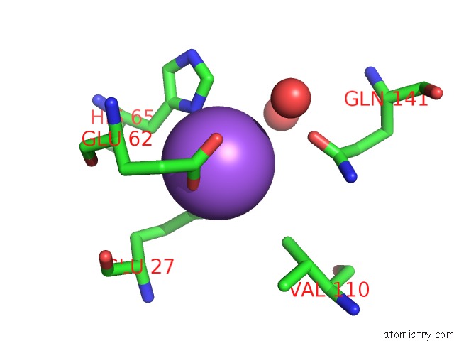
The binding sites of Sodium atom in the 1.15 A Structure of Human Apoferritin Obtained From Titan Mono- Bcor Microscope (pdb code 7a6a). This binding sites where shown within 5.0 Angstroms radius around Sodium atom.In total 32 binding sites of Sodium where determined in the 1.15 A Structure of Human Apoferritin Obtained From Titan Mono- Bcor Microscope, PDB code: 7a6a:
Jump to Sodium binding site number: 1; 2; 3; 4; 5; 6; 7; 8; 9; 10;
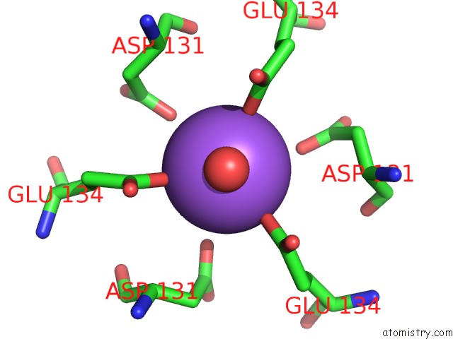
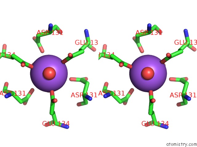
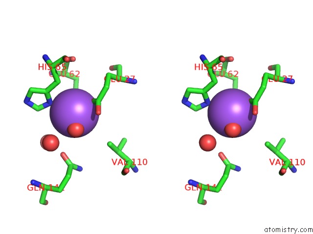
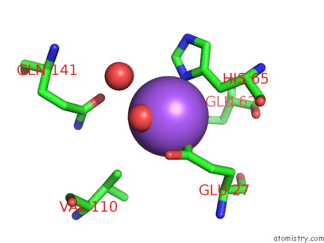
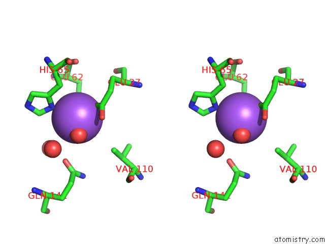
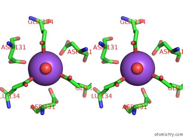
Sodium binding site 1 out of 32 in 7a6a
Go back to
Sodium binding site 1 out
of 32 in the 1.15 A Structure of Human Apoferritin Obtained From Titan Mono- Bcor Microscope

Mono view
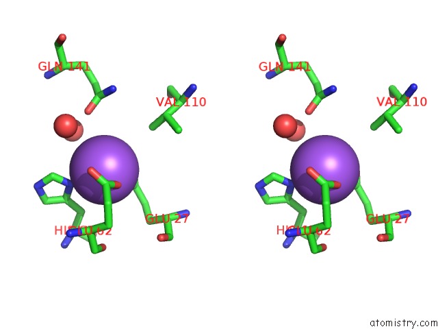
Stereo pair view
Mono view
Stereo pair view
A full contact list of Sodium with other atoms in the Na binding
site number 1 of 1.15 A Structure of Human Apoferritin Obtained From Titan Mono- Bcor Microscope within 5.0Å range:
|
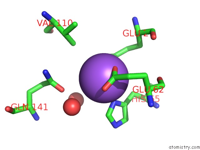
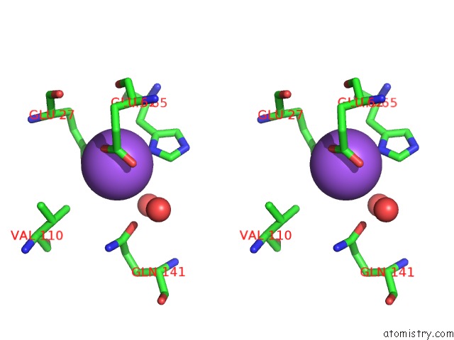
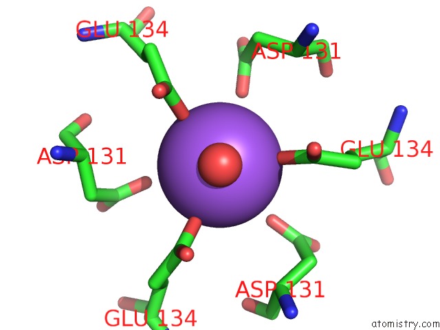
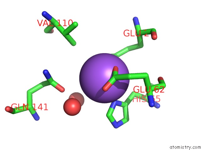
Sodium binding site 2 out of 32 in 7a6a
Go back to
Sodium binding site 2 out
of 32 in the 1.15 A Structure of Human Apoferritin Obtained From Titan Mono- Bcor Microscope
Mono view
Stereo pair view
Mono view
Stereo pair view
A full contact list of Sodium with other atoms in the Na binding
site number 2 of 1.15 A Structure of Human Apoferritin Obtained From Titan Mono- Bcor Microscope within 5.0Å range:
|
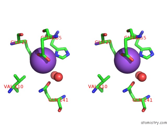
Sodium binding site 3 out of 32 in 7a6a
Go back to
Sodium binding site 3 out
of 32 in the 1.15 A Structure of Human Apoferritin Obtained From Titan Mono- Bcor Microscope
Mono view

Stereo pair view
Mono view
Stereo pair view
A full contact list of Sodium with other atoms in the Na binding
site number 3 of 1.15 A Structure of Human Apoferritin Obtained From Titan Mono- Bcor Microscope within 5.0Å range:
|
Sodium binding site 4 out of 32 in 7a6a
Go back to
Sodium binding site 4 out
of 32 in the 1.15 A Structure of Human Apoferritin Obtained From Titan Mono- Bcor Microscope
Mono view
Stereo pair view
Mono view
Stereo pair view
A full contact list of Sodium with other atoms in the Na binding
site number 4 of 1.15 A Structure of Human Apoferritin Obtained From Titan Mono- Bcor Microscope within 5.0Å range:
|
Sodium binding site 5 out of 32 in 7a6a
Go back to
Sodium binding site 5 out
of 32 in the 1.15 A Structure of Human Apoferritin Obtained From Titan Mono- Bcor Microscope
Mono view
Stereo pair view
Mono view
Stereo pair view
A full contact list of Sodium with other atoms in the Na binding
site number 5 of 1.15 A Structure of Human Apoferritin Obtained From Titan Mono- Bcor Microscope within 5.0Å range:
|
Sodium binding site 6 out of 32 in 7a6a
Go back to
Sodium binding site 6 out
of 32 in the 1.15 A Structure of Human Apoferritin Obtained From Titan Mono- Bcor Microscope

Mono view
Stereo pair view
Mono view
Stereo pair view
A full contact list of Sodium with other atoms in the Na binding
site number 6 of 1.15 A Structure of Human Apoferritin Obtained From Titan Mono- Bcor Microscope within 5.0Å range:
|
Sodium binding site 7 out of 32 in 7a6a
Go back to
Sodium binding site 7 out
of 32 in the 1.15 A Structure of Human Apoferritin Obtained From Titan Mono- Bcor Microscope
Mono view
Stereo pair view
Mono view
Stereo pair view
A full contact list of Sodium with other atoms in the Na binding
site number 7 of 1.15 A Structure of Human Apoferritin Obtained From Titan Mono- Bcor Microscope within 5.0Å range:
|
Sodium binding site 8 out of 32 in 7a6a
Go back to
Sodium binding site 8 out
of 32 in the 1.15 A Structure of Human Apoferritin Obtained From Titan Mono- Bcor Microscope
Mono view
Stereo pair view
Mono view
Stereo pair view
A full contact list of Sodium with other atoms in the Na binding
site number 8 of 1.15 A Structure of Human Apoferritin Obtained From Titan Mono- Bcor Microscope within 5.0Å range:
|
Sodium binding site 9 out of 32 in 7a6a
Go back to
Sodium binding site 9 out
of 32 in the 1.15 A Structure of Human Apoferritin Obtained From Titan Mono- Bcor Microscope

Mono view

Stereo pair view
Mono view
Stereo pair view
A full contact list of Sodium with other atoms in the Na binding
site number 9 of 1.15 A Structure of Human Apoferritin Obtained From Titan Mono- Bcor Microscope within 5.0Å range:
|
Sodium binding site 10 out of 32 in 7a6a
Go back to
Sodium binding site 10 out
of 32 in the 1.15 A Structure of Human Apoferritin Obtained From Titan Mono- Bcor Microscope
Mono view
Stereo pair view
Mono view
Stereo pair view
A full contact list of Sodium with other atoms in the Na binding
site number 10 of 1.15 A Structure of Human Apoferritin Obtained From Titan Mono- Bcor Microscope within 5.0Å range:
|
Reference:
K.M.Yip,
N.Fischer,
A.Chari,
H.Stark.
1.15 A Structure of Human Apoferritin Obtained From Titan Mono- Bcor Microscope To Be Published.
Page generated: Tue Oct 8 15:49:11 2024
Last articles
Fe in 2YXOFe in 2YRS
Fe in 2YXC
Fe in 2YNM
Fe in 2YVJ
Fe in 2YP1
Fe in 2YU2
Fe in 2YU1
Fe in 2YQB
Fe in 2YOO