Sodium »
PDB 6zl4-7a2n »
6ztb »
Sodium in PDB 6ztb: Crystal Structure of Human P-Cadherin EC1_EC2
Protein crystallography data
The structure of Crystal Structure of Human P-Cadherin EC1_EC2, PDB code: 6ztb
was solved by
J.M.Rondeau,
S.Lehmann,
with X-Ray Crystallography technique. A brief refinement statistics is given in the table below:
| Resolution Low / High (Å) | 57.55 / 1.40 |
| Space group | C 1 2 1 |
| Cell size a, b, c (Å), α, β, γ (°) | 120.889, 76.522, 46.208, 90, 107.79, 90 |
| R / Rfree (%) | 19.8 / 21.4 |
Other elements in 6ztb:
The structure of Crystal Structure of Human P-Cadherin EC1_EC2 also contains other interesting chemical elements:
| Calcium | (Ca) | 3 atoms |
Sodium Binding Sites:
The binding sites of Sodium atom in the Crystal Structure of Human P-Cadherin EC1_EC2
(pdb code 6ztb). This binding sites where shown within
5.0 Angstroms radius around Sodium atom.
In total 4 binding sites of Sodium where determined in the Crystal Structure of Human P-Cadherin EC1_EC2, PDB code: 6ztb:
Jump to Sodium binding site number: 1; 2; 3; 4;
In total 4 binding sites of Sodium where determined in the Crystal Structure of Human P-Cadherin EC1_EC2, PDB code: 6ztb:
Jump to Sodium binding site number: 1; 2; 3; 4;
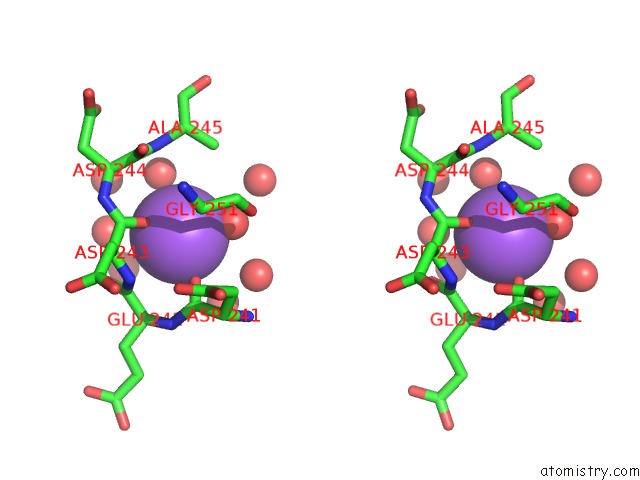
Sodium binding site 1 out of 4 in 6ztb
Go back to
Sodium binding site 1 out
of 4 in the Crystal Structure of Human P-Cadherin EC1_EC2

Mono view
Stereo pair view
Mono view
Stereo pair view
A full contact list of Sodium with other atoms in the Na binding
site number 1 of Crystal Structure of Human P-Cadherin EC1_EC2 within 5.0Å range:
|
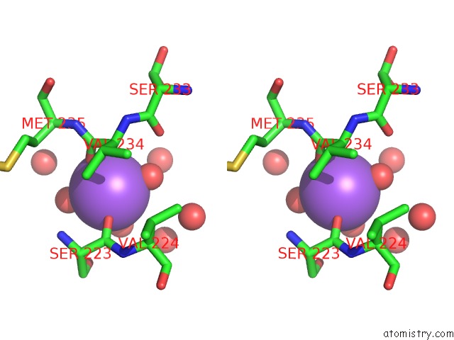
Sodium binding site 2 out of 4 in 6ztb
Go back to
Sodium binding site 2 out
of 4 in the Crystal Structure of Human P-Cadherin EC1_EC2

Mono view
Stereo pair view
Mono view
Stereo pair view
A full contact list of Sodium with other atoms in the Na binding
site number 2 of Crystal Structure of Human P-Cadherin EC1_EC2 within 5.0Å range:
|
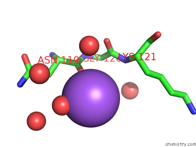
Sodium binding site 3 out of 4 in 6ztb
Go back to
Sodium binding site 3 out
of 4 in the Crystal Structure of Human P-Cadherin EC1_EC2
Mono view

Stereo pair view
Mono view
Stereo pair view
A full contact list of Sodium with other atoms in the Na binding
site number 3 of Crystal Structure of Human P-Cadherin EC1_EC2 within 5.0Å range:
|
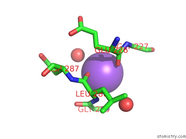
Sodium binding site 4 out of 4 in 6ztb
Go back to
Sodium binding site 4 out
of 4 in the Crystal Structure of Human P-Cadherin EC1_EC2
Mono view

Stereo pair view
Mono view
Stereo pair view
A full contact list of Sodium with other atoms in the Na binding
site number 4 of Crystal Structure of Human P-Cadherin EC1_EC2 within 5.0Å range:
|
Reference:
Q.Sheng,
J.A.D'alessio,
D.L.Menezes,
C.Karim,
Y.Tang,
A.Tam,
S.Clark,
C.Ying,
A.Connor,
K.G.Mansfield,
J.M.Rondeau,
M.Ghoddusi,
F.C.Geyer,
J.Gu,
M.E.Mclaughlin,
R.Newcombe,
G.Elliott,
W.R.Tschantz,
S.Lehmann,
K.Miller,
T.Huber,
K.G.Rendahl,
U.Jeffry,
N.K.Pryer,
E.Lees,
P.Kwon,
J.A.Abraham,
J.S.Damiano,
T.J.Abrams.
PCA062, A P-Cadherin Targeting Antibody-Drug-Conjugate, Displays Potent Anti-Tumor Activity Against P-Cadherin-Expressing Malignancies. Mol.Cancer Ther. 2021.
ISSN: ESSN 1538-8514
PubMed: 33879555
DOI: 10.1158/1535-7163.MCT-20-0708
Page generated: Tue Oct 8 15:40:01 2024
ISSN: ESSN 1538-8514
PubMed: 33879555
DOI: 10.1158/1535-7163.MCT-20-0708
Last articles
Zn in 9MJ5Zn in 9HNW
Zn in 9G0L
Zn in 9FNE
Zn in 9DZN
Zn in 9E0I
Zn in 9D32
Zn in 9DAK
Zn in 8ZXC
Zn in 8ZUF