Sodium in PDB 6z6u: 1.25 A Structure of Human Apoferritin Obtained From Titan Mono-Bcor Microscope
Enzymatic activity of 1.25 A Structure of Human Apoferritin Obtained From Titan Mono-Bcor Microscope
All present enzymatic activity of 1.25 A Structure of Human Apoferritin Obtained From Titan Mono-Bcor Microscope:
1.16.3.1;
Other elements in 6z6u:
The structure of 1.25 A Structure of Human Apoferritin Obtained From Titan Mono-Bcor Microscope also contains other interesting chemical elements:
Sodium Binding Sites:
Pages:
>>> Page 1 <<<
Page 2, Binding sites: 11 -
20;
Page 3, Binding sites: 21 -
24;
Binding sites:
The binding sites of Sodium atom in the 1.25 A Structure of Human Apoferritin Obtained From Titan Mono-Bcor Microscope
(pdb code 6z6u). This binding sites where shown within
5.0 Angstroms radius around Sodium atom.
In total 24 binding sites of Sodium where determined in the
1.25 A Structure of Human Apoferritin Obtained From Titan Mono-Bcor Microscope, PDB code: 6z6u:
Jump to Sodium binding site number:
1;
2;
3;
4;
5;
6;
7;
8;
9;
10;
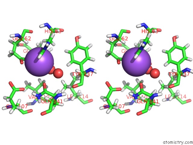
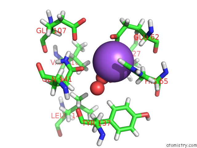
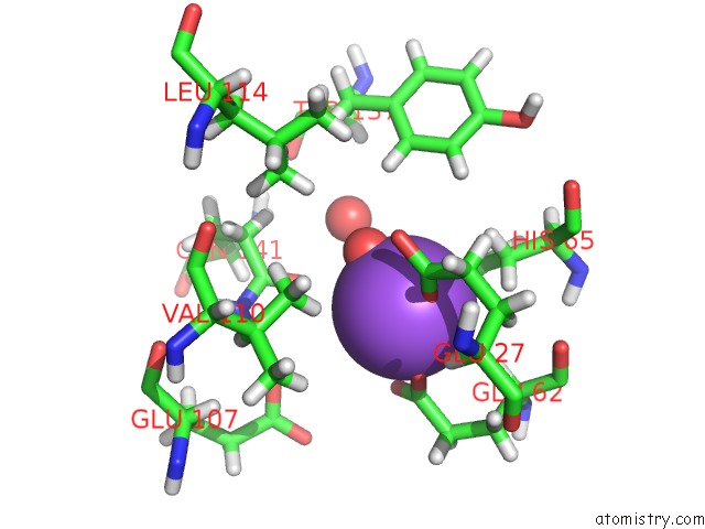
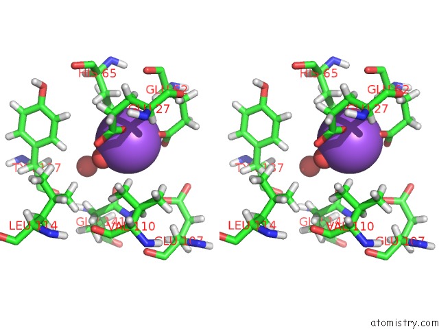
Sodium binding site 1 out
of 24 in 6z6u
Go back to
Sodium Binding Sites List in 6z6u
Sodium binding site 1 out
of 24 in the 1.25 A Structure of Human Apoferritin Obtained From Titan Mono-Bcor Microscope
 Mono view
 Stereo pair view
|
A full contact list of Sodium with other atoms in the Na binding
site number 1 of 1.25 A Structure of Human Apoferritin Obtained From Titan Mono-Bcor Microscope within 5.0Å range:
probe
|
atom
|
residue
|
distance (Å)
|
B
|
Occ
|
A:Na500
b:37.2
occ:1.00
|
HD1
|
A:HIS65
|
1.6
|
37.4
|
0.0
|
OE1
|
A:GLU27
|
2.0
|
37.9
|
1.0
|
O
|
A:HOH610
|
2.1
|
41.3
|
1.0
|
OE1
|
A:GLU62
|
2.2
|
45.1
|
1.0
|
ND1
|
A:HIS65
|
2.4
|
37.4
|
1.0
|
CD
|
A:GLU62
|
2.8
|
38.4
|
1.0
|
OE2
|
A:GLU62
|
2.9
|
56.2
|
1.0
|
CD
|
A:GLU27
|
2.9
|
32.6
|
1.0
|
HB2
|
A:HIS65
|
3.3
|
23.4
|
1.0
|
OE2
|
A:GLU27
|
3.3
|
33.4
|
1.0
|
CE1
|
A:HIS65
|
3.4
|
45.4
|
1.0
|
HG12
|
A:VAL110
|
3.4
|
25.6
|
1.0
|
CG
|
A:HIS65
|
3.4
|
29.7
|
1.0
|
HB3
|
A:HIS65
|
3.5
|
23.4
|
1.0
|
HE1
|
A:HIS65
|
3.6
|
45.4
|
1.0
|
HA
|
A:GLU62
|
3.6
|
19.9
|
1.0
|
CB
|
A:HIS65
|
3.7
|
23.4
|
1.0
|
OE1
|
A:GLN141
|
3.9
|
37.9
|
1.0
|
O
|
A:HOH614
|
4.0
|
42.0
|
1.0
|
CG
|
A:GLU62
|
4.2
|
26.1
|
1.0
|
CG
|
A:GLU27
|
4.2
|
27.2
|
1.0
|
HB3
|
A:GLU27
|
4.3
|
20.8
|
1.0
|
CG1
|
A:VAL110
|
4.3
|
25.6
|
1.0
|
HB3
|
A:GLU62
|
4.5
|
21.8
|
1.0
|
HD2
|
A:TYR137
|
4.5
|
24.7
|
1.0
|
CA
|
A:GLU62
|
4.5
|
19.9
|
1.0
|
HA
|
A:GLU27
|
4.5
|
17.7
|
1.0
|
NE2
|
A:HIS65
|
4.5
|
43.1
|
1.0
|
CD2
|
A:HIS65
|
4.6
|
33.9
|
1.0
|
HG11
|
A:VAL110
|
4.6
|
25.6
|
1.0
|
HG3
|
A:GLU27
|
4.6
|
27.2
|
1.0
|
HE2
|
A:TYR137
|
4.6
|
23.4
|
1.0
|
HG13
|
A:VAL110
|
4.6
|
25.6
|
1.0
|
CB
|
A:GLU62
|
4.6
|
21.8
|
1.0
|
HG2
|
A:GLU62
|
4.7
|
26.1
|
1.0
|
HG3
|
A:GLU62
|
4.7
|
26.1
|
1.0
|
CB
|
A:GLU27
|
4.7
|
20.8
|
1.0
|
CD
|
A:GLN141
|
4.8
|
29.0
|
1.0
|
HG22
|
A:VAL110
|
4.8
|
24.1
|
1.0
|
HG2
|
A:GLU27
|
4.9
|
27.2
|
1.0
|
HD12
|
A:LEU114
|
4.9
|
26.9
|
1.0
|
HE21
|
A:GLN141
|
5.0
|
33.2
|
0.0
|
OE1
|
A:GLU107
|
5.0
|
45.3
|
1.0
|
O
|
A:GLU62
|
5.0
|
19.6
|
1.0
|
|
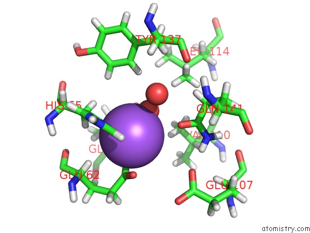
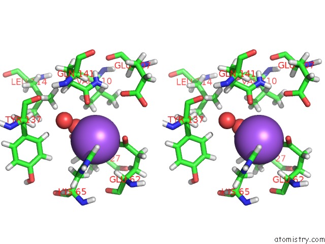
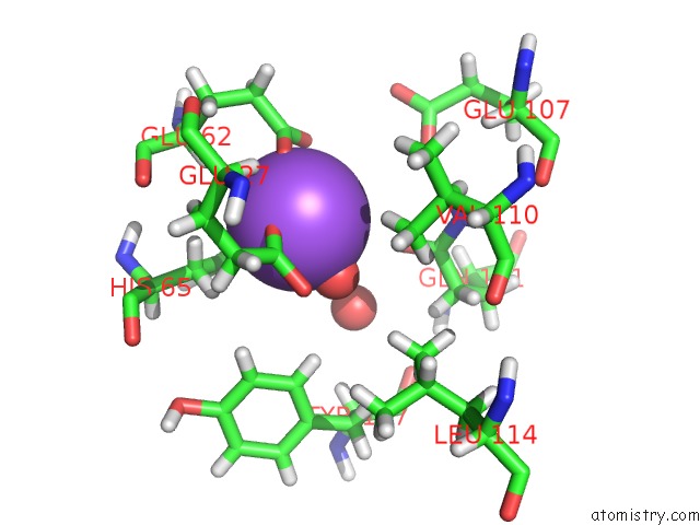
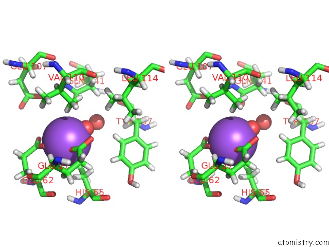
Sodium binding site 2 out
of 24 in 6z6u
Go back to
Sodium Binding Sites List in 6z6u
Sodium binding site 2 out
of 24 in the 1.25 A Structure of Human Apoferritin Obtained From Titan Mono-Bcor Microscope
 Mono view
 Stereo pair view
|
A full contact list of Sodium with other atoms in the Na binding
site number 2 of 1.25 A Structure of Human Apoferritin Obtained From Titan Mono-Bcor Microscope within 5.0Å range:
probe
|
atom
|
residue
|
distance (Å)
|
B
|
Occ
|
K:Na500
b:37.2
occ:1.00
|
HD1
|
K:HIS65
|
1.6
|
37.4
|
0.0
|
OE1
|
K:GLU27
|
2.0
|
37.9
|
1.0
|
O
|
K:HOH610
|
2.1
|
41.3
|
1.0
|
OE1
|
K:GLU62
|
2.2
|
45.1
|
1.0
|
ND1
|
K:HIS65
|
2.4
|
37.4
|
1.0
|
CD
|
K:GLU62
|
2.8
|
38.4
|
1.0
|
OE2
|
K:GLU62
|
2.9
|
56.2
|
1.0
|
CD
|
K:GLU27
|
2.9
|
32.6
|
1.0
|
HB2
|
K:HIS65
|
3.3
|
23.4
|
1.0
|
OE2
|
K:GLU27
|
3.3
|
33.4
|
1.0
|
CE1
|
K:HIS65
|
3.4
|
45.4
|
1.0
|
HG12
|
K:VAL110
|
3.4
|
25.6
|
1.0
|
CG
|
K:HIS65
|
3.4
|
29.7
|
1.0
|
HB3
|
K:HIS65
|
3.5
|
23.4
|
1.0
|
HE1
|
K:HIS65
|
3.6
|
45.4
|
1.0
|
HA
|
K:GLU62
|
3.6
|
19.9
|
1.0
|
CB
|
K:HIS65
|
3.6
|
23.4
|
1.0
|
OE1
|
K:GLN141
|
3.9
|
37.9
|
1.0
|
O
|
K:HOH614
|
4.0
|
42.0
|
1.0
|
CG
|
K:GLU62
|
4.2
|
26.1
|
1.0
|
CG
|
K:GLU27
|
4.2
|
27.2
|
1.0
|
HB3
|
K:GLU27
|
4.3
|
20.8
|
1.0
|
CG1
|
K:VAL110
|
4.3
|
25.6
|
1.0
|
HB3
|
K:GLU62
|
4.5
|
21.8
|
1.0
|
HD2
|
K:TYR137
|
4.5
|
24.7
|
1.0
|
CA
|
K:GLU62
|
4.5
|
19.9
|
1.0
|
HA
|
K:GLU27
|
4.5
|
17.7
|
1.0
|
NE2
|
K:HIS65
|
4.5
|
43.1
|
1.0
|
CD2
|
K:HIS65
|
4.6
|
33.9
|
1.0
|
HG11
|
K:VAL110
|
4.6
|
25.6
|
1.0
|
HG3
|
K:GLU27
|
4.6
|
27.2
|
1.0
|
HE2
|
K:TYR137
|
4.6
|
23.4
|
1.0
|
HG13
|
K:VAL110
|
4.6
|
25.6
|
1.0
|
CB
|
K:GLU62
|
4.6
|
21.8
|
1.0
|
HG2
|
K:GLU62
|
4.7
|
26.1
|
1.0
|
HG3
|
K:GLU62
|
4.7
|
26.1
|
1.0
|
CB
|
K:GLU27
|
4.7
|
20.8
|
1.0
|
CD
|
K:GLN141
|
4.8
|
29.0
|
1.0
|
HG22
|
K:VAL110
|
4.8
|
24.1
|
1.0
|
HG2
|
K:GLU27
|
4.9
|
27.2
|
1.0
|
HD12
|
K:LEU114
|
4.9
|
26.9
|
1.0
|
HE21
|
K:GLN141
|
5.0
|
33.2
|
0.0
|
OE1
|
K:GLU107
|
5.0
|
45.3
|
1.0
|
O
|
K:GLU62
|
5.0
|
19.6
|
1.0
|
|
Sodium binding site 3 out
of 24 in 6z6u
Go back to
Sodium Binding Sites List in 6z6u
Sodium binding site 3 out
of 24 in the 1.25 A Structure of Human Apoferritin Obtained From Titan Mono-Bcor Microscope
 Mono view
 Stereo pair view
|
A full contact list of Sodium with other atoms in the Na binding
site number 3 of 1.25 A Structure of Human Apoferritin Obtained From Titan Mono-Bcor Microscope within 5.0Å range:
probe
|
atom
|
residue
|
distance (Å)
|
B
|
Occ
|
a:Na201
b:37.2
occ:1.00
|
HD1
|
a:HIS65
|
1.6
|
37.4
|
0.0
|
OE1
|
a:GLU27
|
2.0
|
37.9
|
1.0
|
O
|
a:HOH610
|
2.1
|
41.3
|
1.0
|
OE1
|
a:GLU62
|
2.2
|
45.1
|
1.0
|
ND1
|
a:HIS65
|
2.4
|
37.4
|
1.0
|
CD
|
a:GLU62
|
2.8
|
38.4
|
1.0
|
OE2
|
a:GLU62
|
2.9
|
56.2
|
1.0
|
CD
|
a:GLU27
|
2.9
|
32.6
|
1.0
|
HB2
|
a:HIS65
|
3.3
|
23.4
|
1.0
|
OE2
|
a:GLU27
|
3.3
|
33.4
|
1.0
|
CE1
|
a:HIS65
|
3.4
|
45.4
|
1.0
|
HG12
|
a:VAL110
|
3.4
|
25.6
|
1.0
|
CG
|
a:HIS65
|
3.4
|
29.7
|
1.0
|
HB3
|
a:HIS65
|
3.5
|
23.4
|
1.0
|
HE1
|
a:HIS65
|
3.6
|
45.4
|
1.0
|
HA
|
a:GLU62
|
3.6
|
19.9
|
1.0
|
CB
|
a:HIS65
|
3.7
|
23.4
|
1.0
|
OE1
|
a:GLN141
|
3.9
|
37.9
|
1.0
|
O
|
a:HOH613
|
4.0
|
42.0
|
1.0
|
CG
|
a:GLU62
|
4.2
|
26.1
|
1.0
|
CG
|
a:GLU27
|
4.2
|
27.2
|
1.0
|
HB3
|
a:GLU27
|
4.3
|
20.8
|
1.0
|
CG1
|
a:VAL110
|
4.3
|
25.6
|
1.0
|
HB3
|
a:GLU62
|
4.5
|
21.8
|
1.0
|
HD2
|
a:TYR137
|
4.5
|
24.7
|
1.0
|
CA
|
a:GLU62
|
4.5
|
19.9
|
1.0
|
HA
|
a:GLU27
|
4.5
|
17.7
|
1.0
|
NE2
|
a:HIS65
|
4.5
|
43.1
|
1.0
|
CD2
|
a:HIS65
|
4.6
|
33.9
|
1.0
|
HG11
|
a:VAL110
|
4.6
|
25.6
|
1.0
|
HG3
|
a:GLU27
|
4.6
|
27.2
|
1.0
|
HE2
|
a:TYR137
|
4.6
|
23.4
|
1.0
|
HG13
|
a:VAL110
|
4.6
|
25.6
|
1.0
|
CB
|
a:GLU62
|
4.6
|
21.8
|
1.0
|
HG2
|
a:GLU62
|
4.7
|
26.1
|
1.0
|
HG3
|
a:GLU62
|
4.7
|
26.1
|
1.0
|
CB
|
a:GLU27
|
4.7
|
20.8
|
1.0
|
CD
|
a:GLN141
|
4.8
|
29.0
|
1.0
|
HG22
|
a:VAL110
|
4.8
|
24.1
|
1.0
|
HG2
|
a:GLU27
|
4.9
|
27.2
|
1.0
|
HD12
|
a:LEU114
|
4.9
|
26.9
|
1.0
|
HE21
|
a:GLN141
|
5.0
|
33.2
|
0.0
|
OE1
|
a:GLU107
|
5.0
|
45.3
|
1.0
|
O
|
a:GLU62
|
5.0
|
19.6
|
1.0
|
|
Sodium binding site 4 out
of 24 in 6z6u
Go back to
Sodium Binding Sites List in 6z6u
Sodium binding site 4 out
of 24 in the 1.25 A Structure of Human Apoferritin Obtained From Titan Mono-Bcor Microscope
 Mono view
 Stereo pair view
|
A full contact list of Sodium with other atoms in the Na binding
site number 4 of 1.25 A Structure of Human Apoferritin Obtained From Titan Mono-Bcor Microscope within 5.0Å range:
probe
|
atom
|
residue
|
distance (Å)
|
B
|
Occ
|
B:Na500
b:37.2
occ:1.00
|
HD1
|
B:HIS65
|
1.6
|
37.4
|
0.0
|
OE1
|
B:GLU27
|
2.0
|
37.9
|
1.0
|
O
|
B:HOH610
|
2.1
|
41.3
|
1.0
|
OE1
|
B:GLU62
|
2.2
|
45.1
|
1.0
|
ND1
|
B:HIS65
|
2.4
|
37.4
|
1.0
|
CD
|
B:GLU62
|
2.8
|
38.4
|
1.0
|
OE2
|
B:GLU62
|
2.9
|
56.2
|
1.0
|
CD
|
B:GLU27
|
2.9
|
32.6
|
1.0
|
HB2
|
B:HIS65
|
3.3
|
23.4
|
1.0
|
OE2
|
B:GLU27
|
3.3
|
33.4
|
1.0
|
CE1
|
B:HIS65
|
3.4
|
45.4
|
1.0
|
HG12
|
B:VAL110
|
3.4
|
25.6
|
1.0
|
CG
|
B:HIS65
|
3.4
|
29.7
|
1.0
|
HB3
|
B:HIS65
|
3.5
|
23.4
|
1.0
|
HE1
|
B:HIS65
|
3.6
|
45.4
|
1.0
|
HA
|
B:GLU62
|
3.6
|
19.9
|
1.0
|
CB
|
B:HIS65
|
3.7
|
23.4
|
1.0
|
OE1
|
B:GLN141
|
3.9
|
37.9
|
1.0
|
O
|
B:HOH615
|
4.0
|
42.1
|
1.0
|
CG
|
B:GLU62
|
4.2
|
26.1
|
1.0
|
CG
|
B:GLU27
|
4.2
|
27.2
|
1.0
|
HB3
|
B:GLU27
|
4.3
|
20.8
|
1.0
|
CG1
|
B:VAL110
|
4.3
|
25.6
|
1.0
|
HB3
|
B:GLU62
|
4.5
|
21.8
|
1.0
|
HD2
|
B:TYR137
|
4.5
|
24.7
|
1.0
|
CA
|
B:GLU62
|
4.5
|
19.9
|
1.0
|
HA
|
B:GLU27
|
4.5
|
17.7
|
1.0
|
NE2
|
B:HIS65
|
4.5
|
43.1
|
1.0
|
CD2
|
B:HIS65
|
4.6
|
33.9
|
1.0
|
HG11
|
B:VAL110
|
4.6
|
25.6
|
1.0
|
HG3
|
B:GLU27
|
4.6
|
27.2
|
1.0
|
HE2
|
B:TYR137
|
4.6
|
23.4
|
1.0
|
HG13
|
B:VAL110
|
4.6
|
25.6
|
1.0
|
CB
|
B:GLU62
|
4.6
|
21.8
|
1.0
|
HG2
|
B:GLU62
|
4.7
|
26.1
|
1.0
|
HG3
|
B:GLU62
|
4.7
|
26.1
|
1.0
|
CB
|
B:GLU27
|
4.7
|
20.8
|
1.0
|
CD
|
B:GLN141
|
4.8
|
29.0
|
1.0
|
HG22
|
B:VAL110
|
4.8
|
24.1
|
1.0
|
HG2
|
B:GLU27
|
4.9
|
27.2
|
1.0
|
HD12
|
B:LEU114
|
4.9
|
26.9
|
1.0
|
HE21
|
B:GLN141
|
5.0
|
33.2
|
0.0
|
OE1
|
B:GLU107
|
5.0
|
45.3
|
1.0
|
O
|
B:GLU62
|
5.0
|
19.6
|
1.0
|
|
Sodium binding site 5 out
of 24 in 6z6u
Go back to
Sodium Binding Sites List in 6z6u
Sodium binding site 5 out
of 24 in the 1.25 A Structure of Human Apoferritin Obtained From Titan Mono-Bcor Microscope
 Mono view
 Stereo pair view
|
A full contact list of Sodium with other atoms in the Na binding
site number 5 of 1.25 A Structure of Human Apoferritin Obtained From Titan Mono-Bcor Microscope within 5.0Å range:
probe
|
atom
|
residue
|
distance (Å)
|
B
|
Occ
|
E:Na500
b:37.2
occ:1.00
|
HD1
|
E:HIS65
|
1.6
|
37.4
|
0.0
|
OE1
|
E:GLU27
|
2.0
|
37.9
|
1.0
|
O
|
E:HOH610
|
2.1
|
41.3
|
1.0
|
OE1
|
E:GLU62
|
2.2
|
45.1
|
1.0
|
ND1
|
E:HIS65
|
2.4
|
37.4
|
1.0
|
CD
|
E:GLU62
|
2.8
|
38.4
|
1.0
|
OE2
|
E:GLU62
|
2.9
|
56.2
|
1.0
|
CD
|
E:GLU27
|
2.9
|
32.6
|
1.0
|
HB2
|
E:HIS65
|
3.3
|
23.4
|
1.0
|
OE2
|
E:GLU27
|
3.3
|
33.4
|
1.0
|
CE1
|
E:HIS65
|
3.4
|
45.4
|
1.0
|
HG12
|
E:VAL110
|
3.4
|
25.6
|
1.0
|
CG
|
E:HIS65
|
3.4
|
29.7
|
1.0
|
HB3
|
E:HIS65
|
3.5
|
23.4
|
1.0
|
HE1
|
E:HIS65
|
3.6
|
45.4
|
1.0
|
HA
|
E:GLU62
|
3.6
|
19.9
|
1.0
|
CB
|
E:HIS65
|
3.7
|
23.4
|
1.0
|
OE1
|
E:GLN141
|
3.9
|
37.9
|
1.0
|
O
|
E:HOH616
|
4.0
|
42.1
|
1.0
|
CG
|
E:GLU62
|
4.2
|
26.1
|
1.0
|
CG
|
E:GLU27
|
4.2
|
27.2
|
1.0
|
HB3
|
E:GLU27
|
4.3
|
20.8
|
1.0
|
CG1
|
E:VAL110
|
4.3
|
25.6
|
1.0
|
HB3
|
E:GLU62
|
4.5
|
21.8
|
1.0
|
HD2
|
E:TYR137
|
4.5
|
24.7
|
1.0
|
CA
|
E:GLU62
|
4.5
|
19.9
|
1.0
|
HA
|
E:GLU27
|
4.5
|
17.7
|
1.0
|
NE2
|
E:HIS65
|
4.5
|
43.1
|
1.0
|
CD2
|
E:HIS65
|
4.6
|
33.9
|
1.0
|
HG11
|
E:VAL110
|
4.6
|
25.6
|
1.0
|
HE2
|
E:TYR137
|
4.6
|
23.4
|
1.0
|
HG3
|
E:GLU27
|
4.6
|
27.2
|
1.0
|
HG13
|
E:VAL110
|
4.6
|
25.6
|
1.0
|
CB
|
E:GLU62
|
4.6
|
21.8
|
1.0
|
HG2
|
E:GLU62
|
4.7
|
26.1
|
1.0
|
HG3
|
E:GLU62
|
4.7
|
26.1
|
1.0
|
CB
|
E:GLU27
|
4.7
|
20.8
|
1.0
|
CD
|
E:GLN141
|
4.8
|
29.0
|
1.0
|
HG22
|
E:VAL110
|
4.8
|
24.1
|
1.0
|
HG2
|
E:GLU27
|
4.9
|
27.2
|
1.0
|
HD12
|
E:LEU114
|
4.9
|
26.9
|
1.0
|
HE21
|
E:GLN141
|
5.0
|
33.2
|
0.0
|
OE1
|
E:GLU107
|
5.0
|
45.3
|
1.0
|
O
|
E:GLU62
|
5.0
|
19.6
|
1.0
|
|
Sodium binding site 6 out
of 24 in 6z6u
Go back to
Sodium Binding Sites List in 6z6u
Sodium binding site 6 out
of 24 in the 1.25 A Structure of Human Apoferritin Obtained From Titan Mono-Bcor Microscope
 Mono view
 Stereo pair view
|
A full contact list of Sodium with other atoms in the Na binding
site number 6 of 1.25 A Structure of Human Apoferritin Obtained From Titan Mono-Bcor Microscope within 5.0Å range:
probe
|
atom
|
residue
|
distance (Å)
|
B
|
Occ
|
e:Na500
b:37.2
occ:1.00
|
HD1
|
e:HIS65
|
1.6
|
37.4
|
0.0
|
OE1
|
e:GLU27
|
2.0
|
37.9
|
1.0
|
O
|
e:HOH610
|
2.1
|
41.3
|
1.0
|
OE1
|
e:GLU62
|
2.2
|
45.1
|
1.0
|
ND1
|
e:HIS65
|
2.4
|
37.4
|
1.0
|
CD
|
e:GLU62
|
2.8
|
38.4
|
1.0
|
OE2
|
e:GLU62
|
2.9
|
56.2
|
1.0
|
CD
|
e:GLU27
|
2.9
|
32.6
|
1.0
|
HB2
|
e:HIS65
|
3.3
|
23.4
|
1.0
|
OE2
|
e:GLU27
|
3.3
|
33.4
|
1.0
|
CE1
|
e:HIS65
|
3.4
|
45.4
|
1.0
|
HG12
|
e:VAL110
|
3.4
|
25.6
|
1.0
|
CG
|
e:HIS65
|
3.4
|
29.7
|
1.0
|
HB3
|
e:HIS65
|
3.5
|
23.4
|
1.0
|
HE1
|
e:HIS65
|
3.6
|
45.4
|
1.0
|
HA
|
e:GLU62
|
3.6
|
19.9
|
1.0
|
CB
|
e:HIS65
|
3.7
|
23.4
|
1.0
|
OE1
|
e:GLN141
|
3.9
|
37.9
|
1.0
|
O
|
e:HOH613
|
4.0
|
42.0
|
1.0
|
CG
|
e:GLU62
|
4.2
|
26.1
|
1.0
|
CG
|
e:GLU27
|
4.2
|
27.2
|
1.0
|
HB3
|
e:GLU27
|
4.3
|
20.8
|
1.0
|
CG1
|
e:VAL110
|
4.3
|
25.6
|
1.0
|
HB3
|
e:GLU62
|
4.5
|
21.8
|
1.0
|
HD2
|
e:TYR137
|
4.5
|
24.7
|
1.0
|
CA
|
e:GLU62
|
4.5
|
19.9
|
1.0
|
HA
|
e:GLU27
|
4.5
|
17.7
|
1.0
|
NE2
|
e:HIS65
|
4.5
|
43.1
|
1.0
|
CD2
|
e:HIS65
|
4.6
|
33.9
|
1.0
|
HG11
|
e:VAL110
|
4.6
|
25.6
|
1.0
|
HG3
|
e:GLU27
|
4.6
|
27.2
|
1.0
|
HE2
|
e:TYR137
|
4.6
|
23.4
|
1.0
|
HG13
|
e:VAL110
|
4.6
|
25.6
|
1.0
|
CB
|
e:GLU62
|
4.6
|
21.8
|
1.0
|
HG2
|
e:GLU62
|
4.7
|
26.1
|
1.0
|
HG3
|
e:GLU62
|
4.7
|
26.1
|
1.0
|
CB
|
e:GLU27
|
4.7
|
20.8
|
1.0
|
CD
|
e:GLN141
|
4.8
|
29.0
|
1.0
|
HG22
|
e:VAL110
|
4.8
|
24.1
|
1.0
|
HG2
|
e:GLU27
|
4.9
|
27.2
|
1.0
|
HD12
|
e:LEU114
|
4.9
|
26.9
|
1.0
|
HE21
|
e:GLN141
|
5.0
|
33.2
|
0.0
|
OE1
|
e:GLU107
|
5.0
|
45.3
|
1.0
|
O
|
e:GLU62
|
5.0
|
19.6
|
1.0
|
|
Sodium binding site 7 out
of 24 in 6z6u
Go back to
Sodium Binding Sites List in 6z6u
Sodium binding site 7 out
of 24 in the 1.25 A Structure of Human Apoferritin Obtained From Titan Mono-Bcor Microscope
 Mono view
 Stereo pair view
|
A full contact list of Sodium with other atoms in the Na binding
site number 7 of 1.25 A Structure of Human Apoferritin Obtained From Titan Mono-Bcor Microscope within 5.0Å range:
probe
|
atom
|
residue
|
distance (Å)
|
B
|
Occ
|
1:Na500
b:37.2
occ:1.00
|
HD1
|
1:HIS65
|
1.6
|
37.4
|
0.0
|
OE1
|
1:GLU27
|
2.0
|
37.9
|
1.0
|
O
|
1:HOH611
|
2.1
|
41.3
|
1.0
|
OE1
|
1:GLU62
|
2.2
|
45.1
|
1.0
|
ND1
|
1:HIS65
|
2.4
|
37.4
|
1.0
|
CD
|
1:GLU62
|
2.8
|
38.4
|
1.0
|
OE2
|
1:GLU62
|
2.9
|
56.2
|
1.0
|
CD
|
1:GLU27
|
2.9
|
32.6
|
1.0
|
HB2
|
1:HIS65
|
3.3
|
23.4
|
1.0
|
OE2
|
1:GLU27
|
3.3
|
33.4
|
1.0
|
CE1
|
1:HIS65
|
3.4
|
45.4
|
1.0
|
HG12
|
1:VAL110
|
3.4
|
25.6
|
1.0
|
CG
|
1:HIS65
|
3.4
|
29.7
|
1.0
|
HB3
|
1:HIS65
|
3.5
|
23.4
|
1.0
|
HE1
|
1:HIS65
|
3.6
|
45.4
|
1.0
|
HA
|
1:GLU62
|
3.6
|
19.9
|
1.0
|
CB
|
1:HIS65
|
3.7
|
23.4
|
1.0
|
OE1
|
1:GLN141
|
3.9
|
37.9
|
1.0
|
O
|
1:HOH614
|
4.0
|
42.1
|
1.0
|
CG
|
1:GLU62
|
4.2
|
26.1
|
1.0
|
CG
|
1:GLU27
|
4.2
|
27.2
|
1.0
|
HB3
|
1:GLU27
|
4.3
|
20.8
|
1.0
|
CG1
|
1:VAL110
|
4.3
|
25.6
|
1.0
|
HB3
|
1:GLU62
|
4.5
|
21.8
|
1.0
|
HD2
|
1:TYR137
|
4.5
|
24.7
|
1.0
|
CA
|
1:GLU62
|
4.5
|
19.9
|
1.0
|
HA
|
1:GLU27
|
4.5
|
17.7
|
1.0
|
NE2
|
1:HIS65
|
4.5
|
43.1
|
1.0
|
CD2
|
1:HIS65
|
4.6
|
33.9
|
1.0
|
HG11
|
1:VAL110
|
4.6
|
25.6
|
1.0
|
HG3
|
1:GLU27
|
4.6
|
27.2
|
1.0
|
HE2
|
1:TYR137
|
4.6
|
23.4
|
1.0
|
HG13
|
1:VAL110
|
4.6
|
25.6
|
1.0
|
CB
|
1:GLU62
|
4.6
|
21.8
|
1.0
|
HG2
|
1:GLU62
|
4.7
|
26.1
|
1.0
|
HG3
|
1:GLU62
|
4.7
|
26.1
|
1.0
|
CB
|
1:GLU27
|
4.7
|
20.8
|
1.0
|
CD
|
1:GLN141
|
4.8
|
29.0
|
1.0
|
HG22
|
1:VAL110
|
4.8
|
24.1
|
1.0
|
HG2
|
1:GLU27
|
4.9
|
27.2
|
1.0
|
HD12
|
1:LEU114
|
4.9
|
26.9
|
1.0
|
HE21
|
1:GLN141
|
5.0
|
33.2
|
0.0
|
OE1
|
1:GLU107
|
5.0
|
45.3
|
1.0
|
O
|
1:GLU62
|
5.0
|
19.6
|
1.0
|
|
Sodium binding site 8 out
of 24 in 6z6u
Go back to
Sodium Binding Sites List in 6z6u
Sodium binding site 8 out
of 24 in the 1.25 A Structure of Human Apoferritin Obtained From Titan Mono-Bcor Microscope
 Mono view
 Stereo pair view
|
A full contact list of Sodium with other atoms in the Na binding
site number 8 of 1.25 A Structure of Human Apoferritin Obtained From Titan Mono-Bcor Microscope within 5.0Å range:
probe
|
atom
|
residue
|
distance (Å)
|
B
|
Occ
|
r:Na500
b:37.2
occ:1.00
|
HD1
|
r:HIS65
|
1.6
|
37.4
|
0.0
|
OE1
|
r:GLU27
|
2.0
|
37.9
|
1.0
|
O
|
r:HOH611
|
2.1
|
41.3
|
1.0
|
OE1
|
r:GLU62
|
2.2
|
45.1
|
1.0
|
ND1
|
r:HIS65
|
2.4
|
37.4
|
1.0
|
CD
|
r:GLU62
|
2.8
|
38.4
|
1.0
|
OE2
|
r:GLU62
|
2.9
|
56.2
|
1.0
|
CD
|
r:GLU27
|
2.9
|
32.6
|
1.0
|
HB2
|
r:HIS65
|
3.3
|
23.4
|
1.0
|
OE2
|
r:GLU27
|
3.3
|
33.4
|
1.0
|
CE1
|
r:HIS65
|
3.4
|
45.4
|
1.0
|
HG12
|
r:VAL110
|
3.4
|
25.6
|
1.0
|
CG
|
r:HIS65
|
3.4
|
29.7
|
1.0
|
HB3
|
r:HIS65
|
3.5
|
23.4
|
1.0
|
HE1
|
r:HIS65
|
3.6
|
45.4
|
1.0
|
HA
|
r:GLU62
|
3.6
|
19.9
|
1.0
|
CB
|
r:HIS65
|
3.7
|
23.4
|
1.0
|
OE1
|
r:GLN141
|
3.9
|
37.9
|
1.0
|
O
|
r:HOH614
|
4.0
|
42.0
|
1.0
|
CG
|
r:GLU62
|
4.2
|
26.1
|
1.0
|
CG
|
r:GLU27
|
4.2
|
27.2
|
1.0
|
HB3
|
r:GLU27
|
4.3
|
20.8
|
1.0
|
CG1
|
r:VAL110
|
4.3
|
25.6
|
1.0
|
HB3
|
r:GLU62
|
4.5
|
21.8
|
1.0
|
HD2
|
r:TYR137
|
4.5
|
24.7
|
1.0
|
CA
|
r:GLU62
|
4.5
|
19.9
|
1.0
|
HA
|
r:GLU27
|
4.5
|
17.7
|
1.0
|
NE2
|
r:HIS65
|
4.5
|
43.1
|
1.0
|
CD2
|
r:HIS65
|
4.6
|
33.9
|
1.0
|
HG11
|
r:VAL110
|
4.6
|
25.6
|
1.0
|
HG3
|
r:GLU27
|
4.6
|
27.2
|
1.0
|
HE2
|
r:TYR137
|
4.6
|
23.4
|
1.0
|
HG13
|
r:VAL110
|
4.6
|
25.6
|
1.0
|
CB
|
r:GLU62
|
4.6
|
21.8
|
1.0
|
HG2
|
r:GLU62
|
4.7
|
26.1
|
1.0
|
HG3
|
r:GLU62
|
4.7
|
26.1
|
1.0
|
CB
|
r:GLU27
|
4.7
|
20.8
|
1.0
|
CD
|
r:GLN141
|
4.8
|
29.0
|
1.0
|
HG22
|
r:VAL110
|
4.8
|
24.1
|
1.0
|
HG2
|
r:GLU27
|
4.9
|
27.2
|
1.0
|
HD12
|
r:LEU114
|
4.9
|
26.9
|
1.0
|
HE21
|
r:GLN141
|
5.0
|
33.2
|
0.0
|
OE1
|
r:GLU107
|
5.0
|
45.3
|
1.0
|
O
|
r:GLU62
|
5.0
|
19.6
|
1.0
|
|
Sodium binding site 9 out
of 24 in 6z6u
Go back to
Sodium Binding Sites List in 6z6u
Sodium binding site 9 out
of 24 in the 1.25 A Structure of Human Apoferritin Obtained From Titan Mono-Bcor Microscope
 Mono view
 Stereo pair view
|
A full contact list of Sodium with other atoms in the Na binding
site number 9 of 1.25 A Structure of Human Apoferritin Obtained From Titan Mono-Bcor Microscope within 5.0Å range:
probe
|
atom
|
residue
|
distance (Å)
|
B
|
Occ
|
G:Na201
b:37.2
occ:1.00
|
HD1
|
G:HIS65
|
1.6
|
37.4
|
0.0
|
OE1
|
G:GLU27
|
2.0
|
37.9
|
1.0
|
O
|
G:HOH610
|
2.1
|
41.3
|
1.0
|
OE1
|
G:GLU62
|
2.2
|
45.1
|
1.0
|
ND1
|
G:HIS65
|
2.4
|
37.4
|
1.0
|
CD
|
G:GLU62
|
2.8
|
38.4
|
1.0
|
OE2
|
G:GLU62
|
2.9
|
56.2
|
1.0
|
CD
|
G:GLU27
|
2.9
|
32.6
|
1.0
|
HB2
|
G:HIS65
|
3.3
|
23.4
|
1.0
|
OE2
|
G:GLU27
|
3.3
|
33.4
|
1.0
|
CE1
|
G:HIS65
|
3.4
|
45.4
|
1.0
|
HG12
|
G:VAL110
|
3.4
|
25.6
|
1.0
|
CG
|
G:HIS65
|
3.4
|
29.7
|
1.0
|
HB3
|
G:HIS65
|
3.5
|
23.4
|
1.0
|
HE1
|
G:HIS65
|
3.6
|
45.4
|
1.0
|
HA
|
G:GLU62
|
3.6
|
19.9
|
1.0
|
CB
|
G:HIS65
|
3.6
|
23.4
|
1.0
|
OE1
|
G:GLN141
|
3.9
|
37.9
|
1.0
|
O
|
G:HOH614
|
4.0
|
42.1
|
1.0
|
CG
|
G:GLU62
|
4.2
|
26.1
|
1.0
|
CG
|
G:GLU27
|
4.2
|
27.2
|
1.0
|
HB3
|
G:GLU27
|
4.3
|
20.8
|
1.0
|
CG1
|
G:VAL110
|
4.3
|
25.6
|
1.0
|
HB3
|
G:GLU62
|
4.5
|
21.8
|
1.0
|
HD2
|
G:TYR137
|
4.5
|
24.7
|
1.0
|
CA
|
G:GLU62
|
4.5
|
19.9
|
1.0
|
HA
|
G:GLU27
|
4.5
|
17.7
|
1.0
|
NE2
|
G:HIS65
|
4.5
|
43.1
|
1.0
|
CD2
|
G:HIS65
|
4.6
|
33.9
|
1.0
|
HG11
|
G:VAL110
|
4.6
|
25.6
|
1.0
|
HG3
|
G:GLU27
|
4.6
|
27.2
|
1.0
|
HE2
|
G:TYR137
|
4.6
|
23.4
|
1.0
|
HG13
|
G:VAL110
|
4.6
|
25.6
|
1.0
|
CB
|
G:GLU62
|
4.6
|
21.8
|
1.0
|
HG2
|
G:GLU62
|
4.7
|
26.1
|
1.0
|
HG3
|
G:GLU62
|
4.7
|
26.1
|
1.0
|
CB
|
G:GLU27
|
4.7
|
20.8
|
1.0
|
CD
|
G:GLN141
|
4.8
|
29.0
|
1.0
|
HG22
|
G:VAL110
|
4.8
|
24.1
|
1.0
|
HG2
|
G:GLU27
|
4.9
|
27.2
|
1.0
|
HD12
|
G:LEU114
|
4.9
|
26.9
|
1.0
|
HE21
|
G:GLN141
|
5.0
|
33.2
|
0.0
|
OE1
|
G:GLU107
|
5.0
|
45.3
|
1.0
|
O
|
G:GLU62
|
5.0
|
19.6
|
1.0
|
|
Sodium binding site 10 out
of 24 in 6z6u
Go back to
Sodium Binding Sites List in 6z6u
Sodium binding site 10 out
of 24 in the 1.25 A Structure of Human Apoferritin Obtained From Titan Mono-Bcor Microscope
 Mono view
 Stereo pair view
|
A full contact list of Sodium with other atoms in the Na binding
site number 10 of 1.25 A Structure of Human Apoferritin Obtained From Titan Mono-Bcor Microscope within 5.0Å range:
probe
|
atom
|
residue
|
distance (Å)
|
B
|
Occ
|
I:Na500
b:37.2
occ:1.00
|
HD1
|
I:HIS65
|
1.6
|
37.4
|
0.0
|
OE1
|
I:GLU27
|
2.0
|
37.9
|
1.0
|
O
|
I:HOH610
|
2.1
|
41.3
|
1.0
|
OE1
|
I:GLU62
|
2.2
|
45.1
|
1.0
|
ND1
|
I:HIS65
|
2.4
|
37.4
|
1.0
|
CD
|
I:GLU62
|
2.8
|
38.4
|
1.0
|
OE2
|
I:GLU62
|
2.9
|
56.2
|
1.0
|
CD
|
I:GLU27
|
2.9
|
32.6
|
1.0
|
HB2
|
I:HIS65
|
3.3
|
23.4
|
1.0
|
OE2
|
I:GLU27
|
3.3
|
33.4
|
1.0
|
CE1
|
I:HIS65
|
3.4
|
45.4
|
1.0
|
HG12
|
I:VAL110
|
3.4
|
25.6
|
1.0
|
CG
|
I:HIS65
|
3.4
|
29.7
|
1.0
|
HB3
|
I:HIS65
|
3.5
|
23.4
|
1.0
|
HE1
|
I:HIS65
|
3.6
|
45.4
|
1.0
|
HA
|
I:GLU62
|
3.6
|
19.9
|
1.0
|
CB
|
I:HIS65
|
3.7
|
23.4
|
1.0
|
OE1
|
I:GLN141
|
3.9
|
37.9
|
1.0
|
O
|
I:HOH614
|
4.0
|
42.1
|
1.0
|
CG
|
I:GLU62
|
4.2
|
26.1
|
1.0
|
CG
|
I:GLU27
|
4.2
|
27.2
|
1.0
|
HB3
|
I:GLU27
|
4.3
|
20.8
|
1.0
|
CG1
|
I:VAL110
|
4.3
|
25.6
|
1.0
|
HB3
|
I:GLU62
|
4.5
|
21.8
|
1.0
|
HD2
|
I:TYR137
|
4.5
|
24.7
|
1.0
|
CA
|
I:GLU62
|
4.5
|
19.9
|
1.0
|
HA
|
I:GLU27
|
4.5
|
17.7
|
1.0
|
NE2
|
I:HIS65
|
4.5
|
43.1
|
1.0
|
CD2
|
I:HIS65
|
4.6
|
33.9
|
1.0
|
HG11
|
I:VAL110
|
4.6
|
25.6
|
1.0
|
HG3
|
I:GLU27
|
4.6
|
27.2
|
1.0
|
HE2
|
I:TYR137
|
4.6
|
23.4
|
1.0
|
HG13
|
I:VAL110
|
4.6
|
25.6
|
1.0
|
CB
|
I:GLU62
|
4.6
|
21.8
|
1.0
|
HG2
|
I:GLU62
|
4.7
|
26.1
|
1.0
|
HG3
|
I:GLU62
|
4.7
|
26.1
|
1.0
|
CB
|
I:GLU27
|
4.7
|
20.8
|
1.0
|
CD
|
I:GLN141
|
4.8
|
29.0
|
1.0
|
HG22
|
I:VAL110
|
4.8
|
24.1
|
1.0
|
HG2
|
I:GLU27
|
4.9
|
27.2
|
1.0
|
HD12
|
I:LEU114
|
4.9
|
26.9
|
1.0
|
HE21
|
I:GLN141
|
5.0
|
33.2
|
0.0
|
OE1
|
I:GLU107
|
5.0
|
45.3
|
1.0
|
O
|
I:GLU62
|
5.0
|
19.6
|
1.0
|
|
Reference:
K.M.Yip,
N.Fischer,
E.Paknia,
A.Chari,
H.Stark.
1.25 A Structure of Human Apoferritin Obtained From Titan Mono-Bcor Microscope To Be Published.
Page generated: Tue Oct 8 15:21:00 2024
|