Sodium »
PDB 6sfo-6sxg »
6sr3 »
Sodium in PDB 6sr3: X-Ray Pump X-Ray Probe on Lysozyme.Gd Nanocrystals: 62 Fs Time Delay
Enzymatic activity of X-Ray Pump X-Ray Probe on Lysozyme.Gd Nanocrystals: 62 Fs Time Delay
All present enzymatic activity of X-Ray Pump X-Ray Probe on Lysozyme.Gd Nanocrystals: 62 Fs Time Delay:
3.2.1.17;
3.2.1.17;
Protein crystallography data
The structure of X-Ray Pump X-Ray Probe on Lysozyme.Gd Nanocrystals: 62 Fs Time Delay, PDB code: 6sr3
was solved by
M.Kloos,
A.Gorel,
K.Nass,
with X-Ray Crystallography technique. A brief refinement statistics is given in the table below:
| Resolution Low / High (Å) | 22.81 / 2.30 |
| Space group | P 43 21 2 |
| Cell size a, b, c (Å), α, β, γ (°) | 79.000, 79.000, 39.500, 90.00, 90.00, 90.00 |
| R / Rfree (%) | 21.9 / 28.6 |
Other elements in 6sr3:
The structure of X-Ray Pump X-Ray Probe on Lysozyme.Gd Nanocrystals: 62 Fs Time Delay also contains other interesting chemical elements:
| Gadolinium | (Gd) | 2 atoms |
| Chlorine | (Cl) | 3 atoms |
Sodium Binding Sites:
The binding sites of Sodium atom in the X-Ray Pump X-Ray Probe on Lysozyme.Gd Nanocrystals: 62 Fs Time Delay
(pdb code 6sr3). This binding sites where shown within
5.0 Angstroms radius around Sodium atom.
In total only one binding site of Sodium was determined in the X-Ray Pump X-Ray Probe on Lysozyme.Gd Nanocrystals: 62 Fs Time Delay, PDB code: 6sr3:
In total only one binding site of Sodium was determined in the X-Ray Pump X-Ray Probe on Lysozyme.Gd Nanocrystals: 62 Fs Time Delay, PDB code: 6sr3:
Sodium binding site 1 out of 1 in 6sr3
Go back to
Sodium binding site 1 out
of 1 in the X-Ray Pump X-Ray Probe on Lysozyme.Gd Nanocrystals: 62 Fs Time Delay
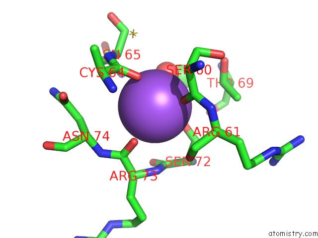
Mono view
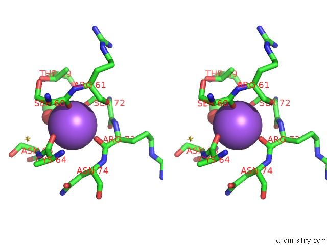
Stereo pair view
Mono view
Stereo pair view
A full contact list of Sodium with other atoms in the Na binding
site number 1 of X-Ray Pump X-Ray Probe on Lysozyme.Gd Nanocrystals: 62 Fs Time Delay within 5.0Å range:
|
Reference:
K.Nass,
A.Gorel,
M.M.Abdullah,
A.V Martin,
M.Kloos,
A.Marinelli,
A.Aquila,
T.R.M.Barends,
F.J.Decker,
R.Bruce Doak,
L.Foucar,
E.Hartmann,
M.Hilpert,
M.S.Hunter,
Z.Jurek,
J.E.Koglin,
A.Kozlov,
A.A.Lutman,
G.N.Kovacs,
C.M.Roome,
R.L.Shoeman,
R.Santra,
H.M.Quiney,
B.Ziaja,
S.Boutet,
I.Schlichting.
Structural Dynamics in Proteins Induced By and Probed with X-Ray Free-Electron Laser Pulses. Nat Commun V. 11 1814 2020.
ISSN: ESSN 2041-1723
PubMed: 32286284
DOI: 10.1038/S41467-020-15610-4
Page generated: Tue Oct 8 13:30:49 2024
ISSN: ESSN 2041-1723
PubMed: 32286284
DOI: 10.1038/S41467-020-15610-4
Last articles
Zn in 9J0NZn in 9J0O
Zn in 9J0P
Zn in 9FJX
Zn in 9EKB
Zn in 9C0F
Zn in 9CAH
Zn in 9CH0
Zn in 9CH3
Zn in 9CH1