Sodium »
PDB 6sfo-6sxg »
6sim »
Sodium in PDB 6sim: Sad Structure of Hen Egg White Lysozyme Recovered By Inverse Beam Geometry Data Collection and Univariate Analysis
Enzymatic activity of Sad Structure of Hen Egg White Lysozyme Recovered By Inverse Beam Geometry Data Collection and Univariate Analysis
All present enzymatic activity of Sad Structure of Hen Egg White Lysozyme Recovered By Inverse Beam Geometry Data Collection and Univariate Analysis:
3.2.1.17;
3.2.1.17;
Protein crystallography data
The structure of Sad Structure of Hen Egg White Lysozyme Recovered By Inverse Beam Geometry Data Collection and Univariate Analysis, PDB code: 6sim
was solved by
M.J.Garcia-Bonete,
G.Katona,
with X-Ray Crystallography technique. A brief refinement statistics is given in the table below:
| Resolution Low / High (Å) | 39.54 / 1.61 |
| Space group | P 43 21 2 |
| Cell size a, b, c (Å), α, β, γ (°) | 79.070, 79.070, 36.980, 90.00, 90.00, 90.00 |
| R / Rfree (%) | 14.6 / 17.4 |
Other elements in 6sim:
The structure of Sad Structure of Hen Egg White Lysozyme Recovered By Inverse Beam Geometry Data Collection and Univariate Analysis also contains other interesting chemical elements:
| Chlorine | (Cl) | 8 atoms |
Sodium Binding Sites:
The binding sites of Sodium atom in the Sad Structure of Hen Egg White Lysozyme Recovered By Inverse Beam Geometry Data Collection and Univariate Analysis
(pdb code 6sim). This binding sites where shown within
5.0 Angstroms radius around Sodium atom.
In total only one binding site of Sodium was determined in the Sad Structure of Hen Egg White Lysozyme Recovered By Inverse Beam Geometry Data Collection and Univariate Analysis, PDB code: 6sim:
In total only one binding site of Sodium was determined in the Sad Structure of Hen Egg White Lysozyme Recovered By Inverse Beam Geometry Data Collection and Univariate Analysis, PDB code: 6sim:
Sodium binding site 1 out of 1 in 6sim
Go back to
Sodium binding site 1 out
of 1 in the Sad Structure of Hen Egg White Lysozyme Recovered By Inverse Beam Geometry Data Collection and Univariate Analysis
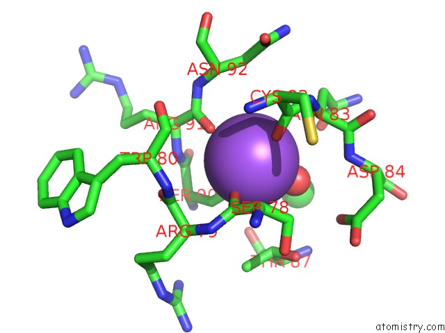
Mono view
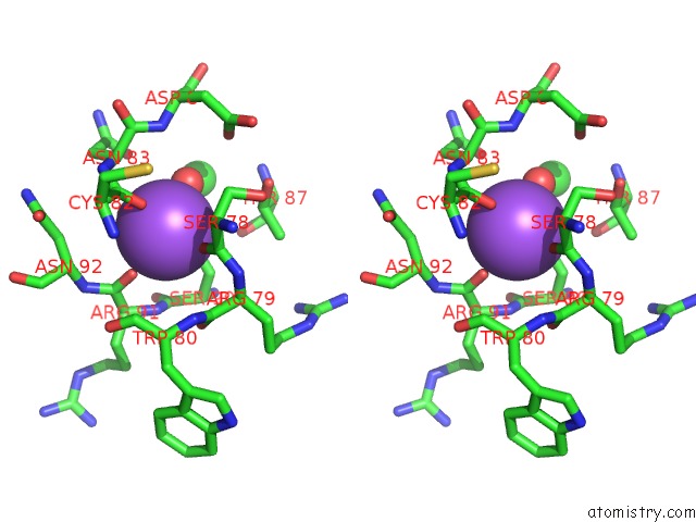
Stereo pair view
Mono view
Stereo pair view
A full contact list of Sodium with other atoms in the Na binding
site number 1 of Sad Structure of Hen Egg White Lysozyme Recovered By Inverse Beam Geometry Data Collection and Univariate Analysis within 5.0Å range:
|
Reference:
M.J.Garcia-Bonete,
G.Katona.
Bayesian Machine Learning Improves Single-Wavelength Anomalous Diffraction Phasing. Acta Crystallogr.,Sect.A V. 75 851 2019.
ISSN: ESSN 2053-2733
PubMed: 31692460
DOI: 10.1107/S2053273319011446
Page generated: Tue Oct 8 13:29:05 2024
ISSN: ESSN 2053-2733
PubMed: 31692460
DOI: 10.1107/S2053273319011446
Last articles
Zn in 9J0NZn in 9J0O
Zn in 9J0P
Zn in 9FJX
Zn in 9EKB
Zn in 9C0F
Zn in 9CAH
Zn in 9CH0
Zn in 9CH3
Zn in 9CH1