Sodium »
PDB 6s08-6sez »
6s0u »
Sodium in PDB 6s0u: The Crystal Structure of Kanamycin B Dioxygenase (Kanj) From Streptomyces Kanamyceticus in Complex with Nickel and 2-Oxoglutarate
Enzymatic activity of The Crystal Structure of Kanamycin B Dioxygenase (Kanj) From Streptomyces Kanamyceticus in Complex with Nickel and 2-Oxoglutarate
All present enzymatic activity of The Crystal Structure of Kanamycin B Dioxygenase (Kanj) From Streptomyces Kanamyceticus in Complex with Nickel and 2-Oxoglutarate:
1.14.11.37;
1.14.11.37;
Protein crystallography data
The structure of The Crystal Structure of Kanamycin B Dioxygenase (Kanj) From Streptomyces Kanamyceticus in Complex with Nickel and 2-Oxoglutarate, PDB code: 6s0u
was solved by
B.Mrugala,
P.J.Porebski,
E.Niedzialkowska,
W.Minor,
T.Borowski,
with X-Ray Crystallography technique. A brief refinement statistics is given in the table below:
| Resolution Low / High (Å) | 45.73 / 2.15 |
| Space group | P 1 21 1 |
| Cell size a, b, c (Å), α, β, γ (°) | 49.825, 183.691, 110.169, 90.00, 96.55, 90.00 |
| R / Rfree (%) | 19.6 / 24.4 |
Other elements in 6s0u:
The structure of The Crystal Structure of Kanamycin B Dioxygenase (Kanj) From Streptomyces Kanamyceticus in Complex with Nickel and 2-Oxoglutarate also contains other interesting chemical elements:
| Nickel | (Ni) | 6 atoms |
| Chlorine | (Cl) | 2 atoms |
Sodium Binding Sites:
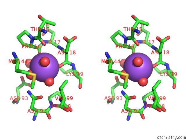
The binding sites of Sodium atom in the The Crystal Structure of Kanamycin B Dioxygenase (Kanj) From Streptomyces Kanamyceticus in Complex with Nickel and 2-Oxoglutarate
(pdb code 6s0u). This binding sites where shown within
5.0 Angstroms radius around Sodium atom.
In total only one binding site of Sodium was determined in the The Crystal Structure of Kanamycin B Dioxygenase (Kanj) From Streptomyces Kanamyceticus in Complex with Nickel and 2-Oxoglutarate, PDB code: 6s0u:
In total only one binding site of Sodium was determined in the The Crystal Structure of Kanamycin B Dioxygenase (Kanj) From Streptomyces Kanamyceticus in Complex with Nickel and 2-Oxoglutarate, PDB code: 6s0u:
Sodium binding site 1 out of 1 in 6s0u
Go back to
Sodium binding site 1 out
of 1 in the The Crystal Structure of Kanamycin B Dioxygenase (Kanj) From Streptomyces Kanamyceticus in Complex with Nickel and 2-Oxoglutarate

Mono view
Stereo pair view
Mono view
Stereo pair view
A full contact list of Sodium with other atoms in the Na binding
site number 1 of The Crystal Structure of Kanamycin B Dioxygenase (Kanj) From Streptomyces Kanamyceticus in Complex with Nickel and 2-Oxoglutarate within 5.0Å range:
|
Reference:
B.Mrugala,
A.Milaczewska,
P.J.Porebski,
E.Niedzialkowska,
M.Guzik,
W.Minor,
T.Borowski.
A Study on the Structure, Mechanism, and Biochemistry of Kanamycin B Dioxygenase (Kanj)-An Enzyme with A Broad Range of Substrates. Febs J. 2020.
ISSN: ISSN 1742-464X
PubMed: 32592631
DOI: 10.1111/FEBS.15462
Page generated: Tue Oct 8 13:22:19 2024
ISSN: ISSN 1742-464X
PubMed: 32592631
DOI: 10.1111/FEBS.15462
Last articles
Zn in 9J0NZn in 9J0O
Zn in 9J0P
Zn in 9FJX
Zn in 9EKB
Zn in 9C0F
Zn in 9CAH
Zn in 9CH0
Zn in 9CH3
Zn in 9CH1