Sodium »
PDB 6rk1-6rzp »
6ru1 »
Sodium in PDB 6ru1: Crystal Structure of Glucuronoyl Esterase From Cerrena Unicolor Inactive S270A Variant in Complex with the Aldouronic Acid UM4X
Protein crystallography data
The structure of Crystal Structure of Glucuronoyl Esterase From Cerrena Unicolor Inactive S270A Variant in Complex with the Aldouronic Acid UM4X, PDB code: 6ru1
was solved by
H.A.Ernst,
C.Mosbech,
A.Langkilde,
P.Westh,
A.Meyer,
J.W.Agger,
S.Larsen,
with X-Ray Crystallography technique. A brief refinement statistics is given in the table below:
| Resolution Low / High (Å) | 42.12 / 1.39 |
| Space group | P 41 21 2 |
| Cell size a, b, c (Å), α, β, γ (°) | 84.240, 84.240, 260.815, 90.00, 90.00, 90.00 |
| R / Rfree (%) | 16.3 / 17.7 |
Sodium Binding Sites:
The binding sites of Sodium atom in the Crystal Structure of Glucuronoyl Esterase From Cerrena Unicolor Inactive S270A Variant in Complex with the Aldouronic Acid UM4X
(pdb code 6ru1). This binding sites where shown within
5.0 Angstroms radius around Sodium atom.
In total only one binding site of Sodium was determined in the Crystal Structure of Glucuronoyl Esterase From Cerrena Unicolor Inactive S270A Variant in Complex with the Aldouronic Acid UM4X, PDB code: 6ru1:
In total only one binding site of Sodium was determined in the Crystal Structure of Glucuronoyl Esterase From Cerrena Unicolor Inactive S270A Variant in Complex with the Aldouronic Acid UM4X, PDB code: 6ru1:
Sodium binding site 1 out of 1 in 6ru1
Go back to
Sodium binding site 1 out
of 1 in the Crystal Structure of Glucuronoyl Esterase From Cerrena Unicolor Inactive S270A Variant in Complex with the Aldouronic Acid UM4X
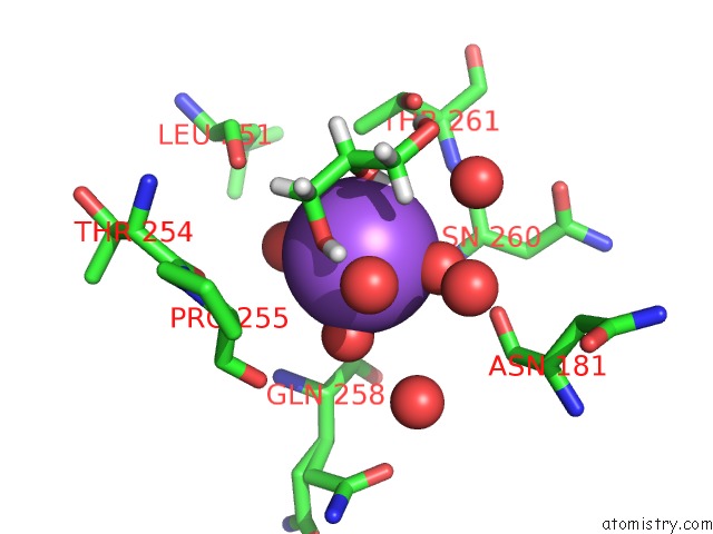
Mono view
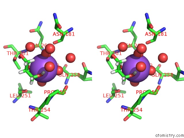
Stereo pair view
Mono view
Stereo pair view
A full contact list of Sodium with other atoms in the Na binding
site number 1 of Crystal Structure of Glucuronoyl Esterase From Cerrena Unicolor Inactive S270A Variant in Complex with the Aldouronic Acid UM4X within 5.0Å range:
|
Reference:
H.A.Ernst,
C.Mosbech,
A.E.Langkilde,
P.Westh,
A.S.Meyer,
J.W.Agger,
S.Larsen.
The Structural Basis of Fungal Glucuronoyl Esterase Activity on Natural Substrates. Nat Commun V. 11 1026 2020.
ISSN: ESSN 2041-1723
PubMed: 32094331
DOI: 10.1038/S41467-020-14833-9
Page generated: Tue Oct 8 13:19:53 2024
ISSN: ESSN 2041-1723
PubMed: 32094331
DOI: 10.1038/S41467-020-14833-9
Last articles
Zn in 9J0NZn in 9J0O
Zn in 9J0P
Zn in 9FJX
Zn in 9EKB
Zn in 9C0F
Zn in 9CAH
Zn in 9CH0
Zn in 9CH3
Zn in 9CH1