Sodium »
PDB 6r7p-6rjn »
6rjn »
Sodium in PDB 6rjn: Crystal Structure of A Fungal Catalase at 2.3 Angstroms
Enzymatic activity of Crystal Structure of A Fungal Catalase at 2.3 Angstroms
All present enzymatic activity of Crystal Structure of A Fungal Catalase at 2.3 Angstroms:
1.11.1.6;
1.11.1.6;
Protein crystallography data
The structure of Crystal Structure of A Fungal Catalase at 2.3 Angstroms, PDB code: 6rjn
was solved by
S.Gomez,
S.Navas-Yuste,
A.M.Payne,
W.Rivera,
M.Lopez-Estepa,
C.Brangbour,
D.Fulla,
J.Juanhuix,
F.J.Fernandez,
M.C.Vega,
with X-Ray Crystallography technique. A brief refinement statistics is given in the table below:
| Resolution Low / High (Å) | 54.67 / 2.30 |
| Space group | P 21 21 2 |
| Cell size a, b, c (Å), α, β, γ (°) | 165.943, 173.690, 96.490, 90.00, 90.00, 90.00 |
| R / Rfree (%) | 14.3 / 19.6 |
Other elements in 6rjn:
The structure of Crystal Structure of A Fungal Catalase at 2.3 Angstroms also contains other interesting chemical elements:
| Potassium | (K) | 7 atoms |
| Iron | (Fe) | 4 atoms |
| Chlorine | (Cl) | 5 atoms |
Sodium Binding Sites:
The binding sites of Sodium atom in the Crystal Structure of A Fungal Catalase at 2.3 Angstroms
(pdb code 6rjn). This binding sites where shown within
5.0 Angstroms radius around Sodium atom.
In total 4 binding sites of Sodium where determined in the Crystal Structure of A Fungal Catalase at 2.3 Angstroms, PDB code: 6rjn:
Jump to Sodium binding site number: 1; 2; 3; 4;
In total 4 binding sites of Sodium where determined in the Crystal Structure of A Fungal Catalase at 2.3 Angstroms, PDB code: 6rjn:
Jump to Sodium binding site number: 1; 2; 3; 4;
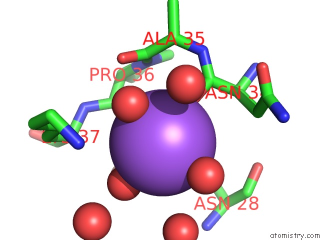
Sodium binding site 1 out of 4 in 6rjn
Go back to
Sodium binding site 1 out
of 4 in the Crystal Structure of A Fungal Catalase at 2.3 Angstroms
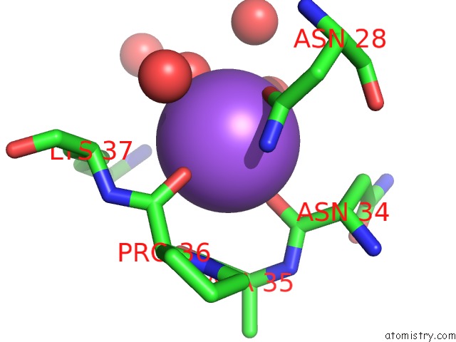
Mono view
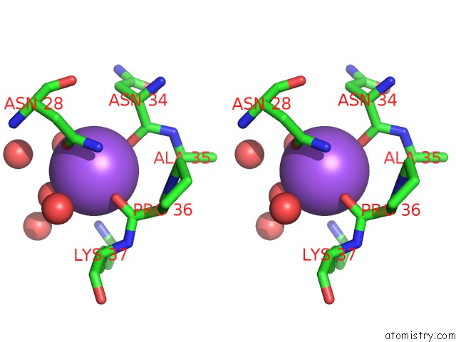
Stereo pair view
Mono view
Stereo pair view
A full contact list of Sodium with other atoms in the Na binding
site number 1 of Crystal Structure of A Fungal Catalase at 2.3 Angstroms within 5.0Å range:
|
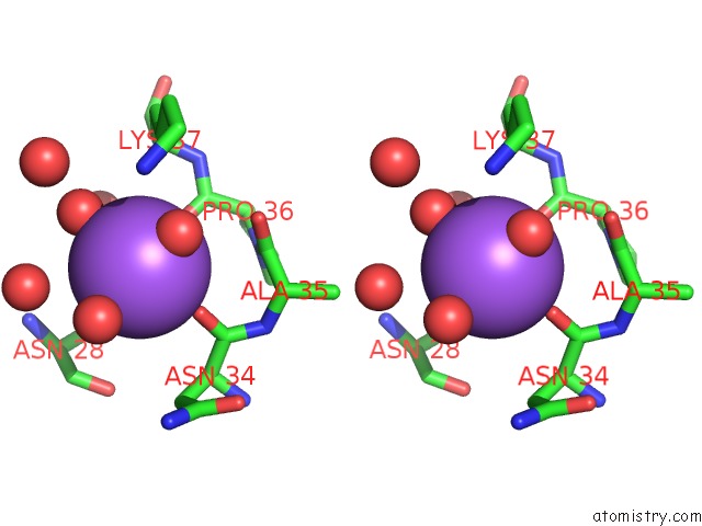
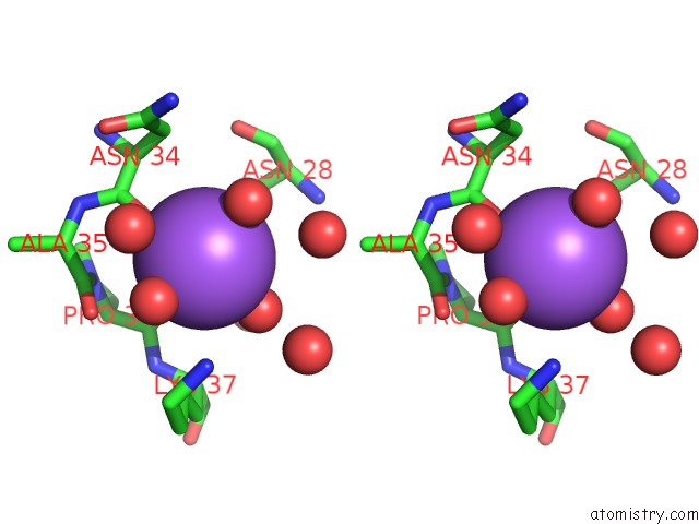
Sodium binding site 2 out of 4 in 6rjn
Go back to
Sodium binding site 2 out
of 4 in the Crystal Structure of A Fungal Catalase at 2.3 Angstroms
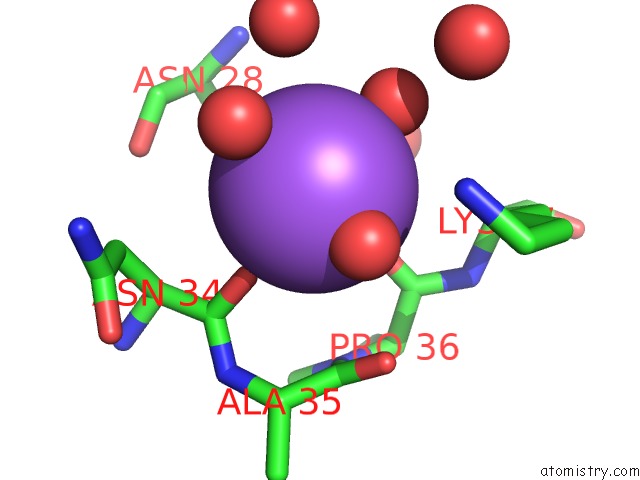
Mono view
Stereo pair view
Mono view
Stereo pair view
A full contact list of Sodium with other atoms in the Na binding
site number 2 of Crystal Structure of A Fungal Catalase at 2.3 Angstroms within 5.0Å range:
|
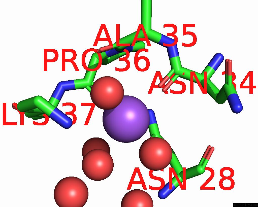
Sodium binding site 3 out of 4 in 6rjn
Go back to
Sodium binding site 3 out
of 4 in the Crystal Structure of A Fungal Catalase at 2.3 Angstroms
Mono view
Stereo pair view
Mono view
Stereo pair view
A full contact list of Sodium with other atoms in the Na binding
site number 3 of Crystal Structure of A Fungal Catalase at 2.3 Angstroms within 5.0Å range:
|
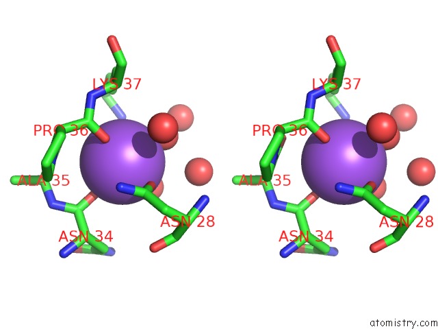
Sodium binding site 4 out of 4 in 6rjn
Go back to
Sodium binding site 4 out
of 4 in the Crystal Structure of A Fungal Catalase at 2.3 Angstroms
Mono view
Stereo pair view
Mono view
Stereo pair view
A full contact list of Sodium with other atoms in the Na binding
site number 4 of Crystal Structure of A Fungal Catalase at 2.3 Angstroms within 5.0Å range:
|
Reference:
S.Gomez,
S.Navas-Yuste,
A.M.Payne,
W.Rivera,
M.Lopez-Estepa,
C.Brangbour,
D.Fulla,
J.Juanhuix,
F.J.Fernandez,
M.C.Vega.
Peroxisomal Catalases From the Yeasts Pichia Pastoris and Kluyveromyces Lactis As Models For Oxidative Damage in Higher Eukaryotes. Free Radic. Biol. Med. V. 141 279 2019.
ISSN: ISSN 1873-4596
PubMed: 31238127
DOI: 10.1016/J.FREERADBIOMED.2019.06.025
Page generated: Tue Oct 8 13:14:20 2024
ISSN: ISSN 1873-4596
PubMed: 31238127
DOI: 10.1016/J.FREERADBIOMED.2019.06.025
Last articles
Cl in 7WM7Cl in 7WLW
Cl in 7WL9
Cl in 7WKZ
Cl in 7WKR
Cl in 7WGP
Cl in 7WK1
Cl in 7WHH
Cl in 7WGT
Cl in 7WF6