Sodium »
PDB 6p9k-6puk »
6phn »
Sodium in PDB 6phn: Crystal Structure of Glucagon Analog Composed of D-Amino Acids with Mono-Stereoinversion at Position 23 (L-VAL23) in Space Group I41 at 1.33 A Resolution
Protein crystallography data
The structure of Crystal Structure of Glucagon Analog Composed of D-Amino Acids with Mono-Stereoinversion at Position 23 (L-VAL23) in Space Group I41 at 1.33 A Resolution, PDB code: 6phn
was solved by
P.A.Mroz,
G.Gonzalez-Gutierrez,
R.D.Dimarchi,
with X-Ray Crystallography technique. A brief refinement statistics is given in the table below:
| Resolution Low / High (Å) | 28.25 / 1.33 |
| Space group | I 41 |
| Cell size a, b, c (Å), α, β, γ (°) | 39.956, 39.956, 38.554, 90.00, 90.00, 90.00 |
| R / Rfree (%) | 16.1 / 19 |
Sodium Binding Sites:
The binding sites of Sodium atom in the Crystal Structure of Glucagon Analog Composed of D-Amino Acids with Mono-Stereoinversion at Position 23 (L-VAL23) in Space Group I41 at 1.33 A Resolution
(pdb code 6phn). This binding sites where shown within
5.0 Angstroms radius around Sodium atom.
In total only one binding site of Sodium was determined in the Crystal Structure of Glucagon Analog Composed of D-Amino Acids with Mono-Stereoinversion at Position 23 (L-VAL23) in Space Group I41 at 1.33 A Resolution, PDB code: 6phn:
In total only one binding site of Sodium was determined in the Crystal Structure of Glucagon Analog Composed of D-Amino Acids with Mono-Stereoinversion at Position 23 (L-VAL23) in Space Group I41 at 1.33 A Resolution, PDB code: 6phn:
Sodium binding site 1 out of 1 in 6phn
Go back to
Sodium binding site 1 out
of 1 in the Crystal Structure of Glucagon Analog Composed of D-Amino Acids with Mono-Stereoinversion at Position 23 (L-VAL23) in Space Group I41 at 1.33 A Resolution
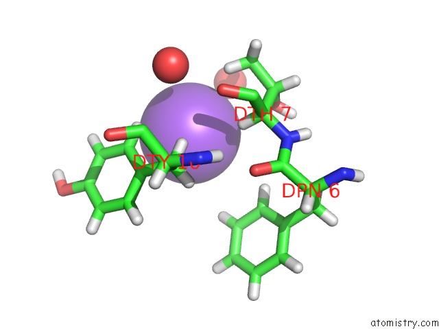
Mono view
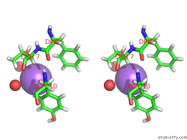
Stereo pair view
Mono view
Stereo pair view
A full contact list of Sodium with other atoms in the Na binding
site number 1 of Crystal Structure of Glucagon Analog Composed of D-Amino Acids with Mono-Stereoinversion at Position 23 (L-VAL23) in Space Group I41 at 1.33 A Resolution within 5.0Å range:
|
Reference:
P.A.Mroz,
G.Gonzalez-Gutierrez,
R.D.Dimarchi.
High Resolution X-Ray Structure of Glucagon and Selected Stereo-Inversed Analogs in Novel Crystallographic Packing Arrangement. To Be Published.
Page generated: Tue Oct 8 12:43:33 2024
Last articles
Fe in 2YXOFe in 2YRS
Fe in 2YXC
Fe in 2YNM
Fe in 2YVJ
Fe in 2YP1
Fe in 2YU2
Fe in 2YU1
Fe in 2YQB
Fe in 2YOO